
Cellular Physiology — MCQs

On this page
Which of the following are force-generating proteins?
Which organ's cells lack the deposition of mitochondria?
Which among the following is the function of cholesterol in the plasma membrane?
Which of the following statements regarding exocytosis is correct?
Exocytosis requires which ion?
Which factor favors transport across a cell membrane?
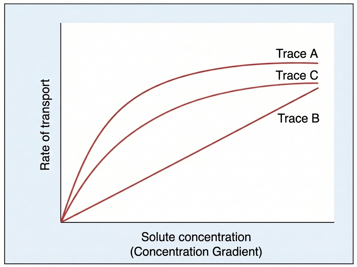
Trace B best describes the kinetics of which of the following events?
Which of the following events does not occur in the M phase of mitosis?
The binding site for which of the following is present on the $\beta$ subunit of the Na+ - K+ pump?
Which are the most common cells to exhibit ameboid locomotion in the human body?
Practice by Chapter
Cell Membrane Structure and Function
Practice Questions
Membrane Transport Proteins
Practice Questions
Cellular Energetics and Metabolism
Practice Questions
Mitochondrial Function
Practice Questions
Cell Volume Regulation
Practice Questions
Cellular Responses to Stress
Practice Questions
Calcium Signaling
Practice Questions
Cell Cycle and Regulation
Practice Questions
Cellular Aging
Practice Questions
Apoptosis and Cell Death
Practice Questions
Want unlimited practice?
Get full access to all questions, explanations, and performance tracking.
Scan to download app