
Cellular Physiology — MCQs

On this page
Across the cell membrane of a skeletal muscle cell in the resting state with a non-electrogenic pump (1 Na+ : 1K+), which of the following statements is true?
All of the following are true about gap junctions except:
All of the following statements about phagocytosis are true, except:
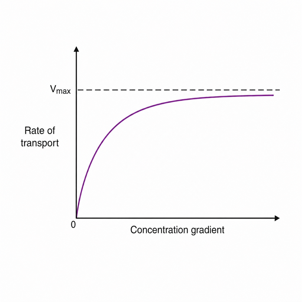
The graph given below shows which of the following transport mechanisms?
Which of the following are force-generating proteins?
Which factor does not affect the diffusion of a substance through the cell membrane?
What is the action of the alpha subunit of a G protein?
What is the extracellular binding site on the Na+ - K+ pump?
Release of neurotransmitter from presynaptic vesicles is an example of?
Active transport and facilitated diffusion share which of the following characteristics?
Practice by Chapter
Cell Membrane Structure and Function
Practice Questions
Membrane Transport Proteins
Practice Questions
Cellular Energetics and Metabolism
Practice Questions
Mitochondrial Function
Practice Questions
Cell Volume Regulation
Practice Questions
Cellular Responses to Stress
Practice Questions
Calcium Signaling
Practice Questions
Cell Cycle and Regulation
Practice Questions
Cellular Aging
Practice Questions
Apoptosis and Cell Death
Practice Questions
Want unlimited practice?
Get full access to all questions, explanations, and performance tracking.
Scan to download app