The electrogenic Na/K ATPase plays a critical role in cellular physiology by?
The endoplasmic reticulum does not participate in which of the following processes?
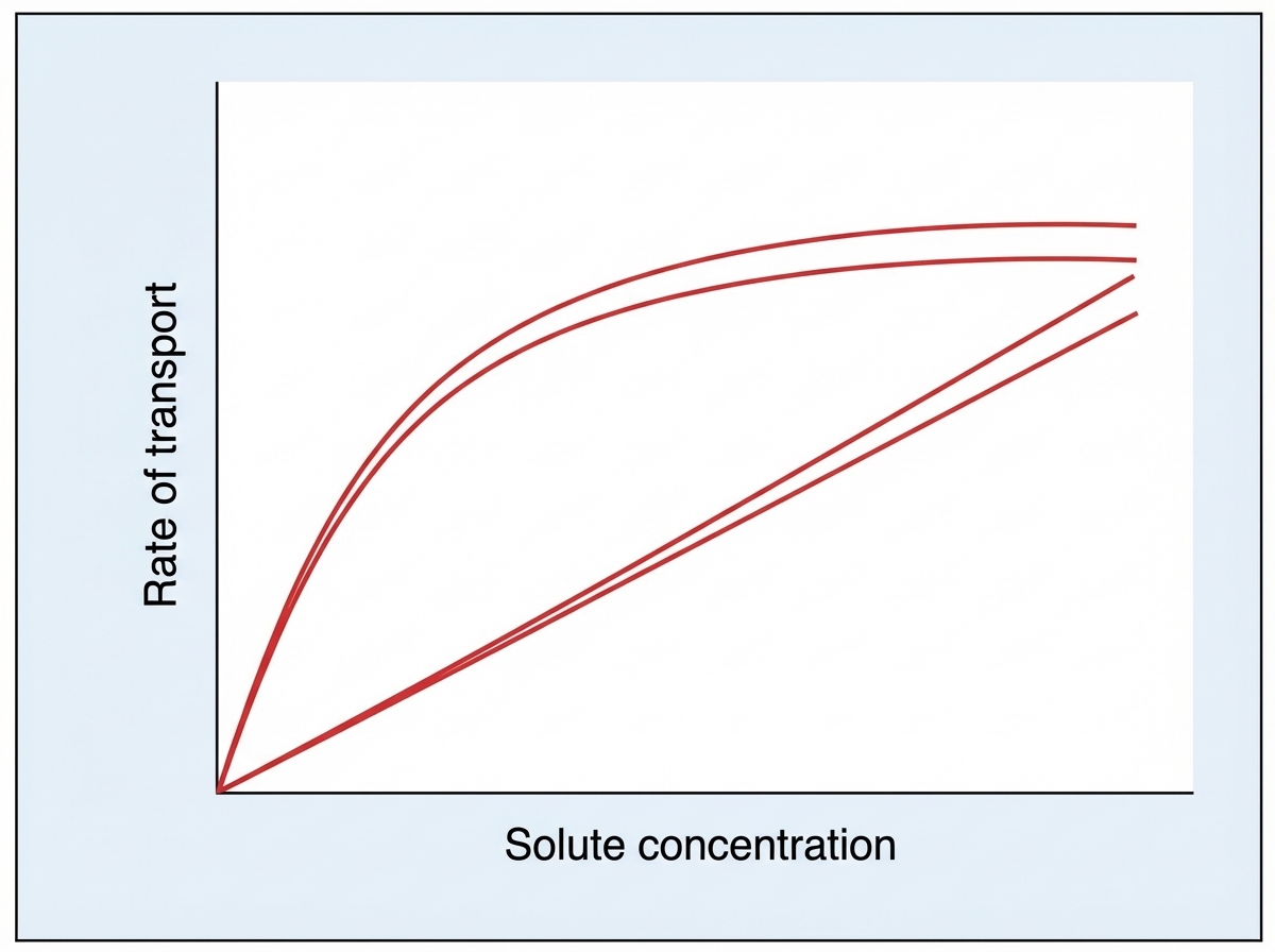
The graph below denotes the transport kinetics across the cell membrane. The solute is:

Which of the following is not a function of the smooth endoplasmic reticulum?
Which of the following is NOT true about active transport?
The cell membrane is permeable to three ions, X, Y, and Z. The equilibrium potentials for ions X and Y are -50 mV and -30 mV, respectively. If the resting membrane potential (RMP) is such that there is no net electrogenic transfer, what is the equilibrium potential for ion Z?
What is the resting membrane potential of a cell?
An increase in extracellular K+ concentration leads to what change in the resting membrane potential (RMP)?
Connexins are associated with which of the following?
Mad cow disease is caused by what?
Explanation: **Explanation:** The **Na+/K+ ATPase pump** is a primary active transporter found in the plasma membrane of all animal cells. It is termed **electrogenic** because it creates a net charge imbalance across the membrane. **Why Option A is Correct:** The pump functions by hydrolyzing one molecule of ATP to provide the energy required to move ions against their respective concentration gradients. In each cycle, it extrudes **3 Na+ ions out** of the cell and brings **2 K+ ions into** the cell. Because three positive charges leave while only two enter, a net deficit of one positive charge occurs inside the cell, contributing directly to the negative resting membrane potential (RMP). **Analysis of Incorrect Options:** * **Option B:** This incorrectly reverses the direction of ion movement. Na+ is always kept at low concentrations intracellularly, while K+ is kept high. * **Options C and D:** These describe the mechanism of **ATP Synthase** (found in mitochondria), which uses electrochemical gradients to *generate* ATP. The Na+/K+ ATPase is a consumer of energy, not a producer. **High-Yield Clinical Pearls for NEET-PG:** * **Inhibitors:** The pump is specifically inhibited by **Cardiac Glycosides** (e.g., Digoxin and Ouabain), which bind to the extracellular alpha-subunit. * **Energy Consumption:** This pump accounts for approximately **33% to 70%** of the total energy expenditure in many cells, particularly neurons. * **Functions:** It is essential for maintaining cell volume (preventing swelling), maintaining the RMP, and providing the Na+ gradient necessary for **secondary active transport** (e.g., SGLT-1 in kidneys/intestines).
Explanation: **Explanation:** The **Endoplasmic Reticulum (ER)** is a multifunctional organelle divided into Rough ER (RER) and Smooth ER (SER). While it plays a central role in biosynthesis and calcium storage, it does not directly participate in the mechanical process of **muscle contraction**. * **Why Option B is correct:** Muscle contraction is a mechanical process occurring in the **sarcomere** through the interaction of actin and myosin filaments (Sliding Filament Theory). While the **Sarcoplasmic Reticulum** (a specialized form of SER) is crucial for storing and releasing calcium ions to *initiate* contraction, the ER itself does not participate in the contractile process. * **Why Option A is incorrect:** The **Rough ER**, studded with ribosomes, is the primary site for the synthesis of secretory, lysosomal, and membrane-bound proteins. * **Why Option C is incorrect:** The ER is the first step in the secretory pathway. It performs **protein sorting** by identifying proteins with specific signal sequences and packaging them into COPII-coated vesicles for transport to the Golgi apparatus. * **Why Option D is incorrect:** **N-linked glycosylation** (the addition of sugar chains to nitrogen atoms of asparagine) begins in the lumen of the Rough ER, making it essential for glycoprotein synthesis. **High-Yield Facts for NEET-PG:** * **Sarcoplasmic Reticulum (SR):** The specialized SER in muscle cells. Its primary function is **Calcium sequestration** via SERCA pumps. * **N-linked vs. O-linked Glycosylation:** N-linked starts in the **ER**; O-linked occurs exclusively in the **Golgi apparatus**. * **Detoxification:** The SER in hepatocytes contains the **Cytochrome P450** system, essential for drug metabolism. * **Protein Quality Control:** The ER contains "chaperones" (e.g., BiP, Calnexin) that ensure proteins are folded correctly before exiting. Misaligned proteins lead to **ER Stress** and the Unfolded Protein Response (UPR).
Explanation: ***Glucose*** - The saturation kinetics curve (Michaelis-Menten-like) with a **plateau at Vmax** is characteristic of **facilitated diffusion** via **GLUT transporters**. - Shows **carrier-mediated transport** where transporters become saturated at high glucose concentrations, unlike simple diffusion. *Oxygen* - Crosses membranes by **simple diffusion** following **Fick's law**, showing a **linear relationship** between concentration and transport rate. - Does not exhibit **saturation kinetics** as it doesn't require specific carriers or channels for transport. *Sodium* - Primarily transported by **active transport** via **Na+/K+-ATPase pumps**, which would show different kinetics than the graph. - Active transport can work **against concentration gradients** and typically shows different curve patterns with energy dependence. *Chloride* - Moves across membranes through **ion channels** or **simple diffusion**, exhibiting **linear kinetics** without saturation. - Does not require **carrier proteins** that would create the saturation pattern shown in the graph.
Explanation: The Smooth Endoplasmic Reticulum (SER) is a multifunctional organelle characterized by the absence of ribosomes on its surface, which dictates its physiological roles. **Why Protein Synthesis is the Correct Answer:** Protein synthesis is the primary function of the **Rough Endoplasmic Reticulum (RER)**. The RER is studded with ribosomes, which are the sites of translation. Proteins synthesized here are typically destined for secretion, incorporation into the cell membrane, or storage in lysosomes. Since the SER lacks ribosomes, it cannot perform protein synthesis. **Analysis of Incorrect Options:** * **Lipid Synthesis:** The SER contains enzymes necessary for the synthesis of phospholipids, cholesterol, and steroid hormones (e.g., testosterone, estrogen, and cortisol). * **Metabolism of Drugs:** The SER in hepatocytes contains the **Cytochrome P450 system**, which is essential for the detoxification of drugs (like phenobarbital) and various toxins. * **Supply of Calcium:** In muscle cells, the SER is specialized as the **Sarcoplasmic Reticulum**, which stores and releases calcium ions ($Ca^{2+}$) to trigger muscle contraction. **High-Yield NEET-PG Pearls:** * **Nissl Bodies:** These are large granules of RER found in neurons; they are responsible for high-rate protein synthesis. * **Sarcoplasmic Reticulum:** A specialized SER in myocytes that regulates excitation-contraction coupling via calcium sequestration. * **Hypertrophy of SER:** Chronic use of certain drugs (e.g., barbiturates) can lead to the hypertrophy of the SER in liver cells due to enzyme induction, contributing to drug tolerance.
Explanation: **Explanation:** Active transport is the movement of molecules or ions across a cell membrane against a concentration or electrochemical gradient. **Why Option D is the correct answer:** Active transport is **not necessarily unidirectional**. While it moves substances against a gradient, many active transport systems are **bidirectional** or involve exchange mechanisms. For example, the **Na⁺-K⁺ ATPase pump** (Primary Active Transport) moves 3 Na⁺ ions out of the cell and 2 K⁺ ions into the cell simultaneously. Similarly, secondary active transport (counter-transport) moves substances in opposite directions. Therefore, labeling it strictly "unidirectional" is physiologically incorrect. **Why the other options are wrong:** * **A. It is saturable:** Since active transport relies on specific membrane proteins, it exhibits **saturation kinetics**. Once all available carrier sites are occupied (Vmax), the rate of transport cannot increase further. * **B. It requires energy:** By definition, active transport requires metabolic energy, usually derived from **ATP hydrolysis** (Primary) or the **stored electrochemical gradient** of another ion (Secondary). * **C. It is a carrier-mediated process:** Unlike simple diffusion, active transport always requires specific **integral membrane proteins** (pumps or transporters) to facilitate the movement of solutes. **High-Yield NEET-PG Pearls:** * **Primary Active Transport:** Examples include Na⁺-K⁺ ATPase, Ca²⁺ ATPase (SERCA pump), and H⁺-K⁺ ATPase (Proton pump in gastric parietal cells). * **Secondary Active Transport:** Uses the sodium gradient created by the Na⁺-K⁺ pump. Examples: **SGLT-1** (Glucose/Na⁺ symport in the gut) and **Na⁺-Ca²⁺ exchanger** (counter-transport). * **Digitalis/Ouabain:** These drugs inhibit the Na⁺-K⁺ ATPase, leading to increased intracellular Na⁺ and subsequently increased intracellular Ca²⁺ via the exchanger, enhancing cardiac contractility.
Explanation: ### Explanation The core concept behind this question is the **Electroneutrality Principle** and the **Chord Conductance Equation**. In a steady-state resting membrane potential (RMP), the sum of all ionic currents must be zero ($I_X + I_Y + I_Z = 0$) for there to be no net electrogenic transfer. The RMP of a cell is determined by the weighted average of the equilibrium potentials ($E$) of all permeant ions, based on their relative conductances ($g$). The formula is: $$V_m = \frac{(g_X \cdot E_X) + (g_Y \cdot E_Y) + (g_Z \cdot E_Z)}{g_{total}}$$ In this specific scenario, the question implies a state of equilibrium where the net movement is zero. For the RMP to be stable and "non-electrogenic" in the context of multiple ions, the RMP must lie within the range of the equilibrium potentials of the involved ions. However, mathematically, if the RMP is not provided, we look at the relationship of the driving forces. For the net transfer to be zero, the positive and negative driving forces must balance out. Given $E_X = -50$ mV and $E_Y = -30$ mV, both ions have negative equilibrium potentials. To achieve a balanced state (especially if the RMP is at a typical physiological level), ion Z must have a significantly different (positive) potential to offset the negative pull of X and Y. Among the choices, **80 mV** is the only value that provides the necessary electrochemical gradient to balance the negative potentials of X and Y. #### Analysis of Incorrect Options: * **Option A (-130) & D (-50):** These are too negative. If all ions had negative equilibrium potentials, the RMP would be strongly negative, and there would be no "opposing" force to reach a steady state without constant energy expenditure. * **Option B (10):** While positive, 10 mV is generally insufficient to balance two significantly negative potentials ( -50 and -30) unless the conductance of Z was disproportionately high. #### High-Yield Clinical Pearls for NEET-PG: * **Nernst Equation:** Calculates the equilibrium potential for a *single* ion. * **Goldman-Hodgkin-Katz (GHK) Equation:** Calculates RMP considering *multiple* ions and their membrane permeabilities. * **Potassium ($K^+$):** The most important determinant of RMP because the resting membrane has the highest permeability to $K^+$. * **Na-K ATPase:** Maintains the concentration gradients but only contributes about -5 to -10 mV directly to the RMP (electrogenic effect).
Explanation: The Resting Membrane Potential (RMP) is the electrical potential difference across the plasma membrane when the cell is in a non-excited state. ### **Explanation of the Correct Option** **Option A** is correct because the RMP is primarily determined by the **selective permeability** of the membrane. At rest, the membrane is significantly more permeable to **K⁺** (via leak channels) than to **Na⁺**. According to the Goldman-Hodgkin-Katz equation, the membrane potential will move closest to the equilibrium potential of the ion with the highest permeability. Since K⁺ efflux occurs more readily, the interior of the cell becomes negative, establishing the RMP. ### **Analysis of Incorrect Options** * **Option B:** Inhibiting the Na⁺/K⁺ ATPase (e.g., with Ouabain) does not immediately drop the RMP to zero. The pump is "electrogenic" but only contributes about **-5 to -10 mV** directly. The majority of the RMP is maintained by passive diffusion through leak channels; it would take time for ionic gradients to dissipate enough for the potential to reach zero. * **Option C:** The RMP is **close to**, but not equal to, the equilibrium potential of K⁺ (-94 mV). Small amounts of Na⁺ influx pull the RMP slightly more positive (typically **-70 to -90 mV**). * **Option D:** The equilibrium potential for Na⁺ is approximately **+60 mV**. The RMP is negative, reflecting the dominance of K⁺ conductance over Na⁺. ### **High-Yield NEET-PG Pearls** * **Goldman Equation:** Used to calculate RMP considering multiple ions and their permeabilities. * **Nernst Equation:** Used to calculate the equilibrium potential for a *single* ion. * **Main Contributor:** The most important factor in *maintaining* the RMP is the **K⁺ leak channels**. * **Role of Na⁺/K⁺ ATPase:** It maintains the concentration gradients (High K⁺ inside, High Na⁺ outside) necessary for the RMP to exist.
Explanation: **Explanation:** The Resting Membrane Potential (RMP) of a cell is primarily determined by the concentration gradient of Potassium ($K^+$) across the cell membrane, as the membrane is highly permeable to $K^+$ at rest. This relationship is governed by the **Nernst Equation**. **1. Why Option A is Correct:** Under normal conditions, $K^+$ concentration is much higher inside the cell (~140 mEq/L) than outside (~4 mEq/L). This steep gradient drives $K^+$ to leak out, leaving the interior electronegative (approx. –70 to –90 mV). When **extracellular $K^+$ increases** (Hyperkalemia), the concentration gradient between the inside and outside of the cell decreases. Consequently, less $K^+$ leaves the cell, and the interior becomes less negative (e.g., moving from –90 mV to –70 mV). In physiological terms, moving toward zero is a **decrease in the magnitude of the RMP** (Depolarization). **2. Why Other Options are Incorrect:** * **Options B & D:** While the RMP does change, the relationship is logarithmic (as per the Nernst Equation), not a fixed linear increase of –10 or –20 mV per 10 mEq/L. Furthermore, an "increase" in RMP usually implies becoming more negative (hyperpolarization), which is the opposite of what occurs in hyperkalemia. * **Option C:** RMP is extremely sensitive to extracellular $K^+$ levels; therefore, "no change" is physiologically impossible. **Clinical Pearls for NEET-PG:** * **Hyperkalemia:** Leads to partial depolarization, making cells initially more excitable, but eventually causes inactivation of $Na^+$ channels, leading to cardiac arrhythmias (Tall T-waves on ECG). * **Hypokalemia:** Increases the concentration gradient, causing $K^+$ to exit the cell more rapidly, leading to **Hyperpolarization** (RMP becomes more negative/increases in magnitude). * **Goldman-Hodgkin-Katz Equation:** Used instead of Nernst when considering multiple ions ($Na^+, K^+, Cl^-$) simultaneously.
Explanation: **Explanation:** **Gap Junctions (The Correct Answer):** Gap junctions are specialized intercellular connections that allow the direct passage of ions and small molecules (up to 1000 Da) between adjacent cells. They are composed of transmembrane proteins called **Connexins**. Six connexin molecules assemble to form a hemichannel known as a **Connexon**. When connexons from two neighboring cells align, they form a complete aqueous channel. These junctions are vital for electrical coupling (e.g., in cardiac muscle and smooth muscle) and chemical signaling. **Why other options are incorrect:** * **Desmosomes (Macula Adherens):** These are "spot welds" that provide mechanical strength to tissues. They utilize **Cadherins** (specifically Desmoglein and Desmocollin) linked to intermediate filaments (keratin). * **Hemidesmosomes:** These anchor the basal surface of epithelial cells to the underlying basement membrane. They primarily involve **Integrins**, not connexins. **High-Yield Clinical Pearls for NEET-PG:** * **Cardiac Physiology:** Gap junctions are the structural basis of the **Intercalated Discs**, allowing the heart to function as a functional syncytium. * **Clinical Correlation:** Mutations in Connexin genes are linked to specific pathologies: * **Connexin 26:** Mutations are the most common cause of congenital non-syndromic **sensorineural deafness**. * **Connexin 32:** Linked to X-linked **Charcot-Marie-Tooth disease**. * **Connexin 46/50:** Associated with congenital **cataracts**. * **Permeability:** Gap junction permeability is regulated by intracellular pH and Ca²⁺ levels (high Ca²⁺ or low pH typically closes the channels).
Explanation: **Explanation:** Mad cow disease, medically known as **Bovine Spongiform Encephalopathy (BSE)**, is caused by **Prions**. Prions are unique infectious agents composed entirely of protein, lacking any nucleic acids (DNA or RNA). The underlying pathophysiology involves the **misfolding** of a normal cellular protein called $PrP^C$ (rich in alpha-helices) into an abnormal, pathological isoform called $PrP^{Sc}$ (rich in beta-pleated sheets). This misfolded protein is resistant to proteases, accumulates in the brain, and induces a chain reaction where it triggers other normal proteins to misfold, leading to neuronal death and a "spongiform" (holey) appearance of brain tissue. **Analysis of Incorrect Options:** * **B. Bacterial infection:** Bacteria are complex organisms with cellular structures and DNA. While they cause many CNS infections (e.g., Meningitis), they are not involved in the pathogenesis of spongiform encephalopathies. * **C. Viral infection:** Viruses consist of genetic material (DNA/RNA) encased in a protein coat. Prions are distinct from viruses because they lack genetic material and are resistant to standard sterilization methods that typically kill viruses (like UV light or boiling). * **D. Spirochete infection:** Spirochetes are a specific group of spiral-shaped bacteria (e.g., *Treponema pallidum*). While Neurosyphilis affects the CNS, it does not cause the protein-aggregation pathology seen in BSE. **High-Yield Clinical Pearls for NEET-PG:** * **Human Variant:** The human form of Mad Cow Disease acquired by consuming contaminated beef is **variant Creutzfeldt-Jakob Disease (vCJD)**. * **Histology:** Characterized by intracytoplasmic vacuoles (spongiform change) and amyloid plaques, notably **without** an inflammatory response. * **Sterilization:** Prions are highly resistant; they require autoclaving at $134^\circ\text{C}$ or the use of strong sodium hydroxide (NaOH) for inactivation. * **Genetics:** The PRNP gene on **Chromosome 20** encodes the prion protein.
Cell Membrane Structure and Function
Practice Questions
Membrane Transport Proteins
Practice Questions
Cellular Energetics and Metabolism
Practice Questions
Mitochondrial Function
Practice Questions
Cell Volume Regulation
Practice Questions
Cellular Responses to Stress
Practice Questions
Calcium Signaling
Practice Questions
Cell Cycle and Regulation
Practice Questions
Cellular Aging
Practice Questions
Apoptosis and Cell Death
Practice Questions
Get full access to all questions, explanations, and performance tracking.
Start For Free