
Cellular Physiology — MCQs

On this page
The plasma membrane of a cell is bounded to the cytoskeleton by which of the following molecules?
Which ion is required for exocytosis?
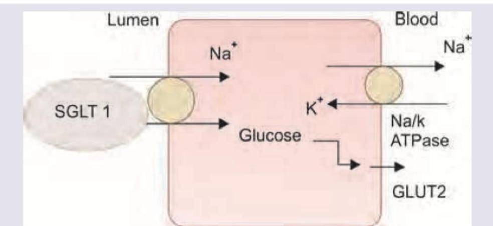
Which of the following statements is true regarding secondary active transport?
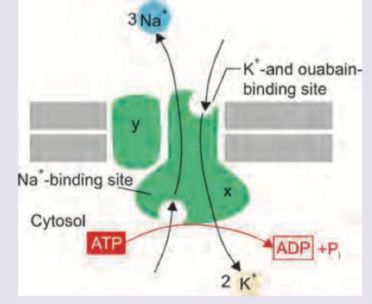
Which of the following is correct about the image shown below?
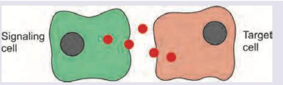
Identify the modality of intercellular communication shown below.
Which of these is true about the highlighted transporter?
Which of the following is not a component of the process shown in the image?
The following graph shows the transport kinetics of a solute transferred across cell membrane. What is the likely mechanism of transport of this solute?

Cystic fibrosis leads to defect in which of the following channels?
Which of the following ion plays a role in exocytosis?
Practice by Chapter
Cell Membrane Structure and Function
Practice Questions
Membrane Transport Proteins
Practice Questions
Cellular Energetics and Metabolism
Practice Questions
Mitochondrial Function
Practice Questions
Cell Volume Regulation
Practice Questions
Cellular Responses to Stress
Practice Questions
Calcium Signaling
Practice Questions
Cell Cycle and Regulation
Practice Questions
Cellular Aging
Practice Questions
Apoptosis and Cell Death
Practice Questions
Want unlimited practice?
Get full access to all questions, explanations, and performance tracking.
Scan to download app