
Cellular Physiology — MCQs

On this page
All of the following statements regarding the Golgi apparatus are true, EXCEPT:
What is true about the Na+-K+ pump?
Which statement is TRUE regarding the Na+/K+ pump?
The electrical potential difference necessary for a single ion to be at equilibrium across a membrane is best described by which equation?
Activation of G-protein regulates all of the following except?
The electrogenic Na/K ATPase plays a critical role in cellular physiology by?
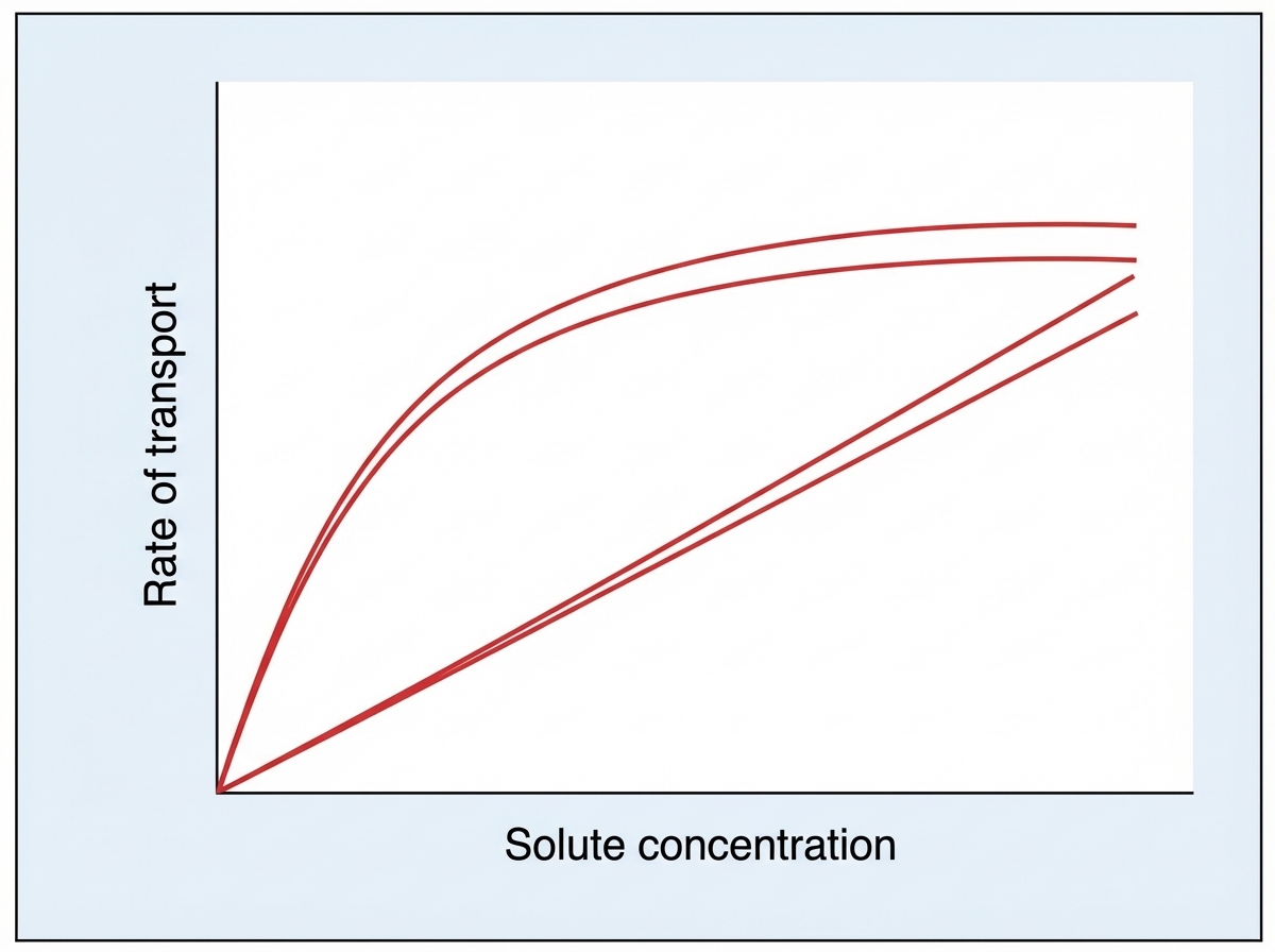
The graph below denotes the transport kinetics across the cell membrane. The solute is:
Which of the following is NOT true about active transport?
A cell membrane is damaged by a needle. How does the repair occur?
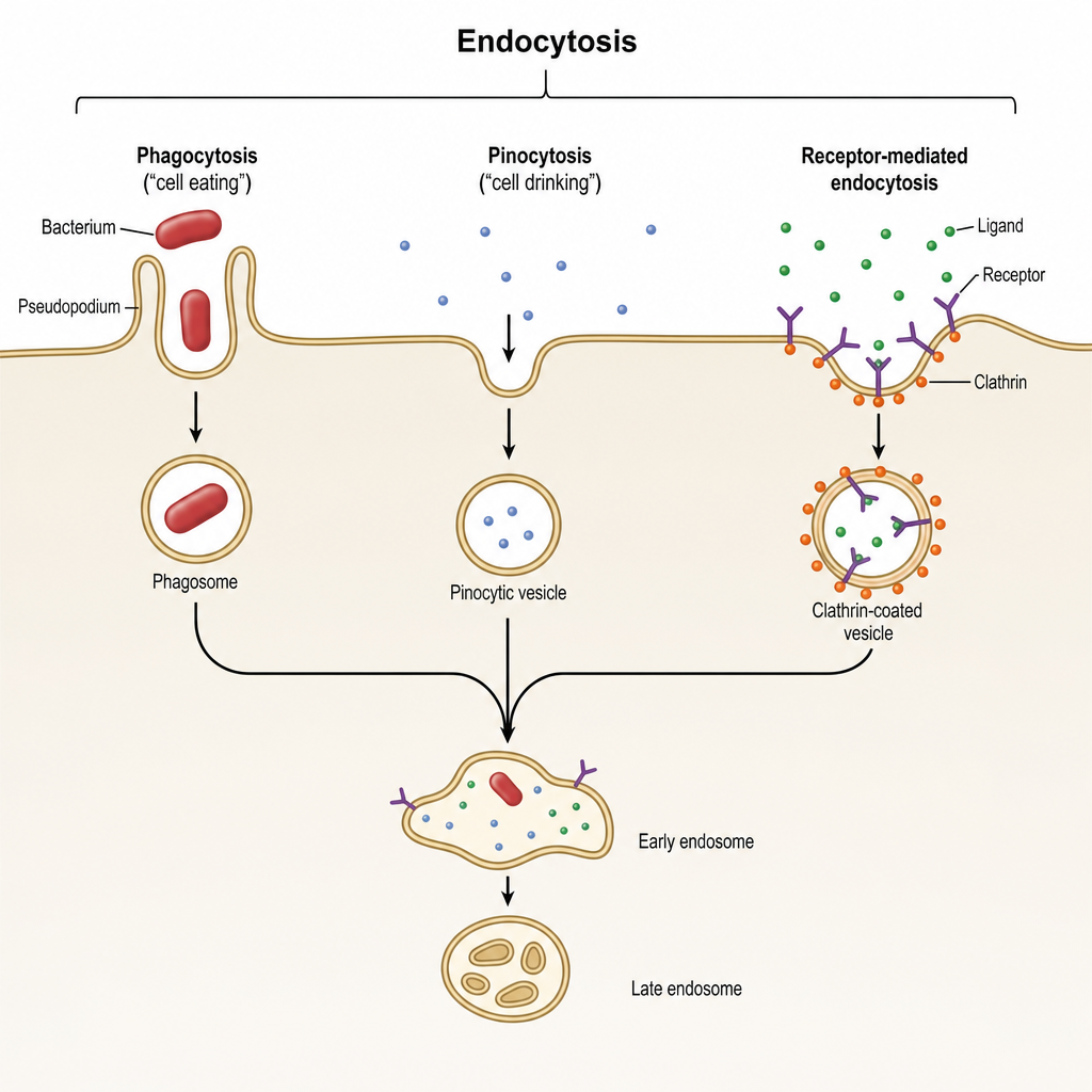
Which statement about the following cellular phenomenon is false?
Practice by Chapter
Cell Membrane Structure and Function
Practice Questions
Membrane Transport Proteins
Practice Questions
Cellular Energetics and Metabolism
Practice Questions
Mitochondrial Function
Practice Questions
Cell Volume Regulation
Practice Questions
Cellular Responses to Stress
Practice Questions
Calcium Signaling
Practice Questions
Cell Cycle and Regulation
Practice Questions
Cellular Aging
Practice Questions
Apoptosis and Cell Death
Practice Questions
Want unlimited practice?
Get full access to all questions, explanations, and performance tracking.
Scan to download app