CO poisoning causes which type of hypoxia?
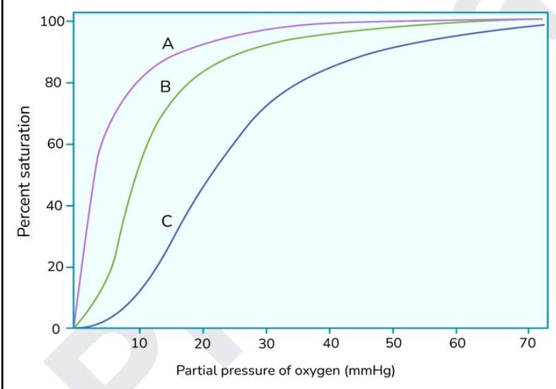
The graph below shows oxygen dissociation curves. What does the curve marked as 'A' indicate?

What is the primary stimulus for erythropoietin production?
What is needed for prothrombin to thrombin conversion?
A pregnant woman is able to transfer oxygen to her fetus because fetal hemoglobin has a greater affinity for oxygen than does adult hemoglobin. Why is the affinity of fetal hemoglobin for oxygen higher?
Decreased O2 carrying capacity and Normal PO2 is a feature of
Oxygen dependent killing is done through
Which of the following cells will increase in case of parasite infection?
Which of the following is not involved in local hemostasis?
All true about interaction of SpO2 reading and methemoglobinemia, except:
Explanation: ***Anemic hypoxia*** - **Carbon monoxide (CO)** binds to **hemoglobin** with an affinity much higher than oxygen, forming **carboxyhemoglobin**. - This effectively reduces the **oxygen-carrying capacity of the blood**, mimicking a severe anemia, despite normal arterial PO2. *Hypoxic hypoxia* - Occurs when there is **insufficient oxygen delivery to the blood** due to low arterial PO2, as seen in high altitudes or respiratory diseases. - In CO poisoning, **arterial PO2 is typically normal**, distinguishing it from hypoxic hypoxia. *Stagnant hypoxia* - Results from **inadequate blood flow** to tissues, leading to reduced oxygen delivery, as observed in heart failure or shock. - CO poisoning primarily affects oxygen transport by hemoglobin, not the **rate of blood flow**. *Histotoxic hypoxia* - Characterized by the **inability of tissues to utilize oxygen** effectively, even when oxygen delivery is adequate, as seen in cyanide poisoning inhibiting cytochrome oxidase. - In CO poisoning, tissues can utilize oxygen; the problem is the **reduced availability of oxygen** from hemoglobin.
Explanation: ***Myoglobin*** - Curve A shows a **hyperbolic oxygen dissociation curve** which is characteristic of myoglobin, indicating high oxygen affinity even at low partial pressures. - Myoglobin has only one heme group, allowing it to bind a single oxygen molecule with high affinity to **store oxygen in muscles**. *Methemoglobin* - Methemoglobin has a **ferric (Fe3+) iron** in its heme group, which cannot bind oxygen, thus reducing the overall oxygen-carrying capacity. - It would typically cause a **left shift** of the remaining functional hemoglobin's dissociation curve due to increased oxygen affinity, but this isn't directly represented as curve A's primary characteristic. *Carboxyhemoglobinemia* - **Carbon monoxide (CO)** binds to hemoglobin with a much higher affinity than oxygen, forming carboxyhemoglobin. - This binding leads to a **left shift** in the oxygen dissociation curve of the remaining functional hemoglobin and a decreased oxygen-carrying capacity, but curve A represents a species with inherently higher oxygen affinity. *Fetal hemoglobin* - Fetal hemoglobin (HbF) has a **higher affinity for oxygen** than adult hemoglobin (HbA), which is represented by a **left-shifted sigmoidal curve** compared to adult hemoglobin. - While it has higher affinity, its curve is still **sigmoidal**, unlike the hyperbolic curve of myoglobin (A).
Explanation: ***Tissue hypoxia*** - Erythropoietin (EPO) production is primarily stimulated by sensing **low oxygen levels** in the kidneys. - This response is crucial for maintaining adequate oxygen delivery to tissues by increasing **red blood cell mass**. *Increased temperature* - An increase in body temperature is a stimulus for processes like **sweating** and **vasodilation**, to regulate body temperature. - It does not directly affect erythropoietin production or red blood cell synthesis. *Decreased blood pressure* - A decrease in blood pressure primarily stimulates the **renin-angiotensin-aldosterone system** and the release of **ADH** to regulate blood volume and pressure. - It does not directly cause an increase in erythropoietin release as its primary function is not related to oxygen sensing. *Decreased plasma proteins* - A decrease in plasma proteins primarily affects **oncotic pressure** and can lead to edema. - It is not a direct stimulus for erythropoietin production.
Explanation: ***Calcium*** - **Calcium ions (Ca2+)** are absolutely essential cofactors for the conversion of **prothrombin to thrombin** in the coagulation cascade. - They are required for the formation and function of the **prothrombinase complex** (Factor Xa + Factor Va + Ca2+ + phospholipid surface). - Calcium binds to **γ-carboxyglutamic acid (Gla) residues** on prothrombin and Factor Xa, enabling them to anchor to phospholipid surfaces where the conversion occurs. - This is why **EDTA and citrate** (calcium chelators) are used as anticoagulants in blood collection tubes. *Magnesium* - **Magnesium** is an important cofactor for numerous enzymatic reactions (e.g., ATP-dependent enzymes, DNA/RNA polymerases). - However, it is **not directly involved** in the prothrombin to thrombin conversion step of the coagulation cascade. - Its primary roles are in DNA synthesis, muscle function, and nerve transmission. *Sodium* - **Sodium** is vital for maintaining fluid balance, osmotic pressure, nerve impulses, and muscle contractions. - It does **not play a direct role** as a cofactor in the prothrombinase complex or prothrombin to thrombin conversion. *Potassium* - **Potassium** is essential for maintaining cell membrane potential, nerve impulses, cardiac function, and muscle contraction. - It is **not a cofactor** for the enzymatic reactions involved in the coagulation cascade.
Explanation: ***Fetal hemoglobin binds 2,3-BPG with fewer ionic bonds than the adult form.*** * **Fetal hemoglobin (HbF)**, composed of two alpha and two gamma subunits, interacts less effectively with **2,3-bisphosphoglycerate (2,3-BPG)** due to a difference in its gamma subunits compared to the beta subunits of **adult hemoglobin (HbA)**. * The reduced binding of 2,3-BPG to HbF stabilizes its **R (relaxed) state**, which has a higher oxygen affinity, facilitating oxygen transfer from the mother to the fetus. *There is less 2,3-BPG in the fetal circulation as compared to maternal circulation* * While 2,3-BPG plays a crucial role in regulating oxygen affinity, the primary reason for **fetal hemoglobin's higher oxygen affinity** is its inherent structural difference that leads to weaker binding of 2,3-BPG, not necessarily the concentration of 2,3-BPG in the fetal circulation. * The **concentration of 2,3-BPG is typically similar or even slightly higher in fetal blood** to enhance oxygen unloading at the tissues, but its effect on HbF is diminished. *The tense form of hemoglobin is more prevalent in the circulation of the fetus* * The **tense form (T-state)** of hemoglobin has a **lower affinity for oxygen**, and its prevalence would lead to reduced oxygen binding, which is contrary to the physiological need of the fetus to extract oxygen from the maternal blood. * **Fetal hemoglobin's higher oxygen affinity** means it spends more time in the **relaxed form (R-state)**, which is responsible for tighter oxygen binding. *The oxygen-binding curve of fetal hemoglobin is shifted to the right.* * An **oxygen-binding curve shifted to the right** indicates a **decreased affinity for oxygen** and would facilitate oxygen unloading, not oxygen loading. * For fetal hemoglobin to effectively extract oxygen from maternal blood, its **oxygen-binding curve must be shifted to the left**, signifying a higher oxygen affinity.
Explanation: ***Anemic hypoxia*** - This condition is characterized by a **reduced number of circulating red blood cells** or a **low hemoglobin concentration**, leading to a decreased capacity to transport oxygen to tissues. - While the overall oxygen-carrying capacity is diminished, the **partial pressure of oxygen (PO2)** in the arterial blood remains normal because the lungs are still efficiently oxygenating the existing hemoglobin. *Hypoxic hypoxia* - This type of hypoxia involves a **decreased partial pressure of oxygen (PO2)** in the arterial blood, often due to conditions affecting lung function or environmental factors (e.g., high altitude). - While there is a reduced amount of oxygen available for transport, the **oxygen-carrying capacity** of the blood itself is not inherently impaired. *Histotoxic hypoxia* - In histotoxic hypoxia, the **cellular machinery responsible for oxygen utilization is impaired**, typically due to toxins like cyanide. - Both the **PO2** and the **oxygen-carrying capacity** of the blood are usually normal, but the tissues cannot effectively use the delivered oxygen. *Stagnant hypoxia* - Also known as **ischemic hypoxia**, this occurs when there is inadequate blood flow to the tissues, leading to insufficient oxygen delivery despite normal **PO2** and **oxygen-carrying capacity**. - This is often seen in conditions like **heart failure** or **localized arterial obstruction**.
Explanation: ***NADPH oxidase*** - **NADPH oxidase** is the enzyme responsible for the **respiratory burst**, producing **superoxide radicals (O2-)** which are crucial for oxygen-dependent killing by phagocytes. - This enzyme converts **molecular oxygen** into highly reactive **superoxide** by reducing NADP+ using NADPH. *Superoxide dismutase* - **Superoxide dismutase (SOD)** neutralizes **superoxide radicals** by converting them into hydrogen peroxide, acting as an antioxidant defense mechanism, rather than a killing mechanism. - While it deals with reactive oxygen species, its role is to protect the cell from oxidative damage, not to generate products for microbial killing. *Glutathione peroxidase* - **Glutathione peroxidase** is an antioxidant enzyme that reduces **hydrogen peroxide** and organic hydroperoxides to water using **reduced glutathione**, thus protecting cells from oxidative stress. - It does not directly produce reactive oxygen species for microbial killing but rather detoxifies them. *Catalase* - **Catalase** converts **hydrogen peroxide** into water and oxygen, functioning to protect cells from the damaging effects of hydrogen peroxide. - Like superoxide dismutase and glutathione peroxidase, its primary role is protective against reactive oxygen species, not in generating them for pathogen eradication.
Explanation: ***Eosinophil*** - **Eosinophils** play a crucial role in the immune response against **parasitic infections**, particularly helminths. - They release cytotoxic granules containing **major basic protein**, **eosinophil cationic protein**, and other mediators that damage the parasites. *Lymphocyte* - **Lymphocytes** are primarily involved in adaptive immunity and are crucial for fighting viral infections and certain bacterial infections, but their increase is not a primary marker for parasitic infections. - While T-helper cells (a type of lymphocyte) can activate eosinophils, a direct increase in total lymphocytes is not the hallmark of parasitic infections. *Basophil* - **Basophils** are involved in allergic reactions and chronic inflammation, releasing histamine and other mediators. - While they can be activated during some parasitic infections, their increase is not as prominent or specific as that of eosinophils. *Neutrophil* - **Neutrophils** are the most abundant white blood cells and are the primary responders to acute bacterial infections and inflammation. - They are less effective against parasitic infections, which often require specialized immune responses.
Explanation: ***Vitamin K*** - **Vitamin K** is crucial for the synthesis of **clotting factors (II, VII, IX, X, protein C, protein S)** in the liver, which are part of the **coagulation cascade** (secondary hemostasis). - Its role is systemic, affecting the production of factors involved in blood coagulation throughout the body, rather than directly participating in the *local* events at the site of injury. *Fibrinogen* - **Fibrinogen** is a soluble protein that is converted to **insoluble fibrin** by thrombin, forming the meshwork of the **blood clot** at the site of injury. - This conversion is a critical local event in strengthening the primary platelet plug during hemostasis. *Collagen* - Exposed **collagen** in the damaged vessel wall directly activates **platelets** (leading to their adhesion and aggregation) and also initiates the **intrinsic pathway** of the coagulation cascade. - It provides the initial signal and a crucial binding site for platelets, essential for forming the primary hemostatic plug. *Calcium* - **Calcium ions (Ca2+)** are essential cofactors for several steps in the **coagulation cascade**, including the activation of various clotting factors (e.g., factors X, IX, VIII, V, and prothrombin). - Its presence is required for the proper functioning of enzyme complexes that form on platelet surfaces, facilitating the generation of thrombin and fibrin at the local injury site.
Explanation: ***Correct: Does not get affected in Methemoglobinemia*** - This statement is **FALSE**, making it the correct answer for this EXCEPT question - **Methemoglobinemia significantly affects SpO2 readings** due to MetHb's optical properties - Pulse oximeters cannot distinguish methemoglobin from oxyhemoglobin and deoxyhemoglobin, leading to **inaccurate measurements** - As MetHb levels rise, the SpO2 reading tends to **plateau around 85%** regardless of true oxygen saturation *Incorrect: Increase in MetHb produces an overestimation when true SpO2 <85%* - This statement is TRUE - When **actual SpO2 is below 85%** and MetHb is elevated, the pulse oximeter reads **higher than the true value** (overestimation) - This occurs because MetHb absorption characteristics cause the reading to gravitate toward 85% *Incorrect: MetHb absorbs red and infrared wavelength of light in a 1:1 ratio* - This statement is TRUE - **Methemoglobin** has similar extinction coefficients at both **660 nm (red)** and **940 nm (infrared)** wavelengths - This 1:1 absorption ratio corresponds to an SpO2 reading of approximately **85%** on conventional pulse oximeters - This is why SpO2 readings plateau at 85% in methemoglobinemia regardless of true saturation *Incorrect: Increase in MetHb produces an underestimation when true SpO2 >85%* - This statement is TRUE - When **actual SpO2 is above 85%** and MetHb is elevated, the pulse oximeter reads **lower than the true value** (underestimation) - The reading is pulled down toward the 85% plateau created by MetHb's absorption characteristics
Composition and Functions of Blood
Practice Questions
Erythrocytes and Hemoglobin
Practice Questions
Leukocytes and Immune Function
Practice Questions
Platelets and Hemostasis
Practice Questions
Blood Groups and Transfusion
Practice Questions
Coagulation and Fibrinolysis
Practice Questions
Hematopoiesis
Practice Questions
Innate Immunity
Practice Questions
Adaptive Immunity
Practice Questions
Immunological Memory and Tolerance
Practice Questions
Get full access to all questions, explanations, and performance tracking.
Start For Free