A 35-year-old woman with menorrhagia presents with pallor, fatigue, and koilonychia. Laboratory findings show hemoglobin 8.2 g/dL, MCV 68 fL, serum iron 25 μg/dL, TIBC 450 μg/dL, and serum ferritin 10 ng/mL. Which of the following best explains the increased TIBC in this patient?
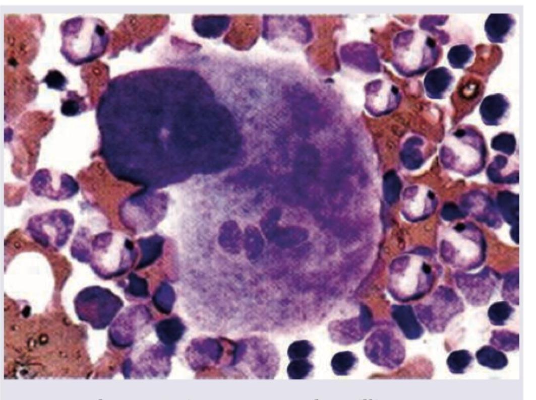
A macrophage engulfs different cells as shown in the image. This is known as?

The least marked function of a human spleen is
Which of the following is X-linked dominant trait?
The genetic inheritance of Haemophilia is
Stored blood which has been preserved in a blood bank is deficient in which of the following coagulation factors?
The removal of malarial parasites from the blood is called
Which one of the following factors is NOT involved in the pathogenesis of Systemic inflammatory response syndrome (SIRS)?
The immunoglobulins that can be transported across the placenta include:
What is the ratio of T cells to B cells in a healthy adult?
Explanation: ***Upregulation of transferrin synthesis by the liver in response to iron deficiency*** - In **Iron Deficiency Anemia (IDA)**, depleted iron stores signal the liver to increase the production of **transferrin** as a compensatory mechanism to maximize iron scavenging and transport from whatever sources are available. - **TIBC (Total Iron Binding Capacity)** directly reflects the number of available iron-binding sites on transferrin, so increased transferrin synthesis leads to **elevated TIBC** (450 μg/dL in this patient). - This is the primary mechanism explaining high TIBC in iron deficiency states. *Increased transferrin degradation in iron overload state* - In **iron overload** conditions (e.g., hemochromatosis), serum iron and ferritin are elevated, and the liver **decreases** (not increases) transferrin synthesis to limit further iron binding and absorption. - Iron overload states are associated with **low TIBC** due to reduced transferrin production, contradicting this patient's presentation of low iron and high TIBC. *Increased erythropoietin production stimulating iron mobilization* - While **erythropoietin (EPO)** production does increase in anemia to stimulate erythropoiesis, EPO does not directly regulate **transferrin synthesis** or TIBC. - EPO acts on bone marrow erythroid progenitors to increase red cell production, but the increase in TIBC is specifically mediated by hepatic transferrin synthesis in response to iron deficiency signals, not EPO. *Decreased hepcidin production leading to increased iron absorption* - **Hepcidin** levels are indeed **decreased** in iron deficiency, which facilitates increased intestinal iron absorption and iron release from macrophages. - However, decreased hepcidin primarily affects iron absorption and mobilization, not the synthesis of transferrin itself, which is the direct determinant of TIBC. - The increased TIBC is due to hepatic upregulation of transferrin production, not hepcidin regulation.
Explanation: ***Emperipolesis*** - The image shows a large cell (likely a macrophage or megakaryocyte) containing **intact various blood cells** within its cytoplasm without signs of degeneration. - **Emperipolesis** is specifically defined as the **active penetration of one cell by another**, where both the engulfed and engulfing cells remain viable. *Phagocytosis* - **Phagocytosis** involves the ingestion and subsequent **destruction or degradation** of foreign particles, microorganisms, or cellular debris. - The cells within the macrophage in the image appear **morphologically intact** and not in a state of degradation. *Killing* - **Killing** implies the process by which a cell actively destroys another cell, often through mechanisms like **apoptosis or necrosis**. - There are **no morphological features** in the image to suggest that the engulfed cells are being actively killed or are undergoing degeneration. *Cytotoxicity* - **Cytotoxicity** refers to the ability of certain immune cells (e.g., cytotoxic T lymphocytes, NK cells) to **kill target cells**. - This process usually involves specific recognition and induction of target cell death, which is not what is depicted by the presence of intact cells within another cell.
Explanation: ***Reservoir Function*** - In **humans**, the spleen stores only a **minimal amount of blood** (~30-40 ml of RBCs), unlike in animals like dogs and horses where it serves as a major blood reservoir - While it does store some **platelets and monocytes**, the reservoir function is the **least marked** among the spleen's primary functions in humans - This function is relatively insignificant compared to the spleen's other critical roles *Filter Function* - The spleen is a **major blood filter**, removing **old and damaged red blood cells**, bacteria, and cellular debris - Removes approximately **20-30 ml of aged RBCs daily** through selective filtration in the red pulp - This is one of the **most important** and highly marked functions of the spleen *Immune Function* - The spleen is a **major secondary lymphoid organ** containing **25% of the body's lymphocytes** - Produces **antibodies** (IgM and IgG) and responds to blood-borne antigens - Critical for fighting **encapsulated organisms** (Streptococcus pneumoniae, Haemophilus influenzae, Neisseria meningitidis) - Post-splenectomy patients face risk of **OPSI** (overwhelming post-splenectomy infection), highlighting its crucial immune role *Pitting Function* - The spleen performs the unique **pitting function** by selectively removing intracellular inclusions (**Howell-Jolly bodies, Pappenheimer bodies, Heinz bodies**) from RBCs without destroying the entire cell - This specialized function helps maintain RBC quality and is particularly evident in splenectomized patients who show these inclusions in peripheral blood
Explanation: ***Vitamin D resistant rickets*** - This condition is characterized by **impaired kidney reabsorption of phosphate** and **defective bone mineralization**, leading to rickets, and it is inherited in an **X-linked dominant pattern**. - In X-linked dominant traits, an affected father will pass the trait to **all his daughters**, but none of his sons. An affected mother has a 50% chance of passing the trait to **each child**, regardless of sex. *Colour blindness* - **Color blindness** is an **X-linked recessive** disorder, meaning it is more common in males. - Affected individuals have **difficulty distinguishing certain colors**, usually red and green. *Haemophilia-A* - **Hemophilia A** is an **X-linked recessive** disorder caused by a deficiency in **Factor VIII**, leading to uncontrolled bleeding. - Males are predominantly affected, while females are typically carriers. *Duchenne muscular dystrophy* - **Duchenne muscular dystrophy** is an **X-linked recessive** disorder characterized by progressive muscle degeneration and weakness due to a mutation in the **dystrophin gene**. - It primarily affects males, with symptoms often appearing in early childhood.
Explanation: ***Sex-linked recessive*** - Hemophilia, specifically hemophilia A and B, is inherited in an **X-linked recessive** pattern. - Males, having only one X chromosome, are predominantly affected, as a single copy of the mutated gene on the X chromosome leads to the condition. *Autosomal recessive* - Autosomal recessive disorders require **two copies** of the mutated gene (one from each parent) to manifest, and affect males and females equally. - This inheritance pattern does not explain the male predominance and carrier status in females seen in hemophilia. *Autosomal dominant* - Autosomal dominant disorders require only **one copy** of the mutated gene to cause the disease, and also affect males and females equally. - This pattern would mean an affected parent has a 50% chance of passing the condition to each child, which is not characteristic of hemophilia. *Sex-linked dominant* - Sex-linked dominant disorders would affect both males and females, with affected fathers passing the trait to **all their daughters** but none of their sons. - This pattern is not consistent with the inheritance of hemophilia, where affected mothers can pass it to their sons.
Explanation: ***V and VIII*** - **Factors V and VIII** are **labile coagulation factors** that degrade quickly during blood storage, making them deficient in stored blood. - This lability is due to their protein structure, which is sensitive to breakdown at refrigerated temperatures over time. *IX and X* - **Factors IX and X** are relatively stable and **vitamin K-dependent factors** that maintain their activity well in stored blood. - They are not significantly depleted during standard blood bank storage periods. *II and VII* - **Factors II (prothrombin) and VII** are also **vitamin K-dependent factors** known for their stability in stored blood. - Their levels remain largely preserved for typical blood storage durations. *II only* - **Factor II (prothrombin)** is a **stable, vitamin K-dependent factor**, and its levels are well-preserved in stored blood. - Therefore, stating only factor II is deficient is incorrect, as it is one of the more stable factors.
Explanation: ***Phagocytosis*** - **Phagocytosis** is the process by which specialized cells, such as **macrophages** and **neutrophils**, engulf and digest foreign particles, including malarial parasites in the blood. - This is a crucial immune mechanism for clearing infected red blood cells and free parasites from the body. - Phagocytosis occurs primarily in the **spleen, liver, and bone marrow**, where macrophages recognize and destroy parasitized RBCs. *Binding* - **Binding** refers to the initial attachment of parasites or infected cells to host cells or tissues. - While binding is a step in the parasitic lifecycle and influences sequestration, it is not the process of *removal* from the blood. *Culling* - **Culling** refers to the selective removal of damaged, aged, or parasitized red blood cells by the spleen. - In malaria, the spleen does "cull" infected RBCs through mechanical filtration and phagocytic clearance. - However, **phagocytosis** is the broader immunological term that describes the cellular mechanism of engulfment and destruction, making it the more appropriate general answer. *Sequestration* - **Sequestration** is the process by which malarial parasites (specifically *Plasmodium falciparum*) in infected red blood cells adhere to the endothelium of capillaries and venules, especially in vital organs. - This mechanism allows parasites to **avoid clearance** by the spleen, representing a process of *retention* or *hiding* rather than removal from the circulation.
Explanation: ***Microvascular occlusion*** - While microvascular occlusion (including microthrombi formation) does occur in SIRS, it is generally considered a **secondary phenomenon or consequence** rather than a **primary initiating pathogenic mechanism**. - It develops as a result of **endothelial dysfunction, platelet activation, and coagulation cascade activation** triggered by the primary inflammatory mediators. - More characteristically associated with **disseminated intravascular coagulation (DIC)**, which is a complication of severe SIRS/sepsis rather than an initiating factor. - The primary pathogenic drivers initiate the cascade, while microvascular occlusion represents a downstream effect contributing to organ dysfunction. *Free radical production* - **Free radicals** (reactive oxygen species and reactive nitrogen species) are directly produced by activated inflammatory cells and damaged tissues. - They cause **oxidative stress**, leading to direct cellular damage, lipid peroxidation, and increased vascular permeability. - Free radical production is a **key pathogenic mechanism** amplifying the inflammatory response and tissue injury in SIRS. *Increased cytokine production* - **Pro-inflammatory cytokines** (TNF-α, IL-1, IL-6, IL-8) are the **central mediators** of SIRS pathogenesis. - They are released early in response to inflammatory stimuli (infection, trauma, burns, pancreatitis). - These cytokines trigger the **systemic inflammatory cascade**, causing fever, vasodilation, capillary leak, leukocyte activation, and acute phase response. - Represent the **primary pathogenic mechanism** driving SIRS. *Abnormal nitric oxide synthesis* - **Excessive nitric oxide (NO) production** by inducible nitric oxide synthase (iNOS) is a **direct pathogenic mechanism** in SIRS. - Leads to **inappropriate vasodilation**, contributing to the **distributive shock** and **refractory hypotension** seen in severe SIRS and septic shock. - NO also impairs vascular responsiveness to vasoconstrictors and contributes to **myocardial depression**. - This abnormal synthesis is a **primary factor** in the hemodynamic instability of SIRS.
Explanation: ***IgG only*** - **IgG** is the only class of immunoglobulin that can actively be transported across the **placental barrier** from mother to fetus, providing passive immunity. - This transport is mediated by specialized receptors (FcRn) on placental cells, ensuring the fetus receives protection against pathogens. *IgA only* - **IgA** is primarily found in **mucosal secretions** (e.g., breast milk, tears, saliva) and plays a crucial role in mucosal immunity. - It does not significantly cross the placenta and therefore does not contribute to fetal immunity in utero. *Neither IgG nor IgA* - This statement is incorrect because **IgG** is well-established as being transported across the placenta. - Such an absence of maternal antibodies would leave the fetus highly vulnerable to infections during development and early life. *Both IgG and IgA* - While **IgG** readily crosses the placenta, **IgA** does not, making this option incorrect. - The primary route for IgA transfer to the infant is through **breast milk** after birth, providing crucial immunity for the newborn's gastrointestinal tract.
Explanation: ***3:1*** - In healthy adults, a typical peripheral blood sample shows a **predominance of T cells** over B cells. - This ratio reflects the differing roles and distributions of these lymphocytes; **T cells are more numerous** in circulation. *1:3* - This ratio would indicate **more B cells than T cells**, which is not typical for peripheral blood in a healthy individual. - An inversion of the usual T:B cell ratio could suggest certain disease states, such as specific **lymphoid malignancies**. *1:1* - While possible in some specific lymphoid tissues, a **1:1 ratio is not the standard** for circulating T and B cells in healthy adults. - This ratio would represent a **significantly higher proportion of B cells** than usually found in peripheral blood. *1:2* - This ratio implies **twice as many B cells as T cells**, which is significantly skewed compared to normal physiological levels in peripheral blood. - Such a high proportion of B cells is typically observed in states of **B-cell proliferation** or specific immune dysregulation.
Composition and Functions of Blood
Practice Questions
Erythrocytes and Hemoglobin
Practice Questions
Leukocytes and Immune Function
Practice Questions
Platelets and Hemostasis
Practice Questions
Blood Groups and Transfusion
Practice Questions
Coagulation and Fibrinolysis
Practice Questions
Hematopoiesis
Practice Questions
Innate Immunity
Practice Questions
Adaptive Immunity
Practice Questions
Immunological Memory and Tolerance
Practice Questions
Get full access to all questions, explanations, and performance tracking.
Start For Free