A 5-year-old child presented with recurrent respiratory infections with thickened sputum. Chest X-ray showed bronchial wall thickening. He has been suffering from steatorrhea since birth. Which of the following is the first differential diagnosis?
Which of the following diagnoses can cause stridor in infants? 1. Laryngomalacia 2. Congenital hemangioma 3. Subglottic stenosis 4. Recurrent respiratory papillomatosis (RRP)
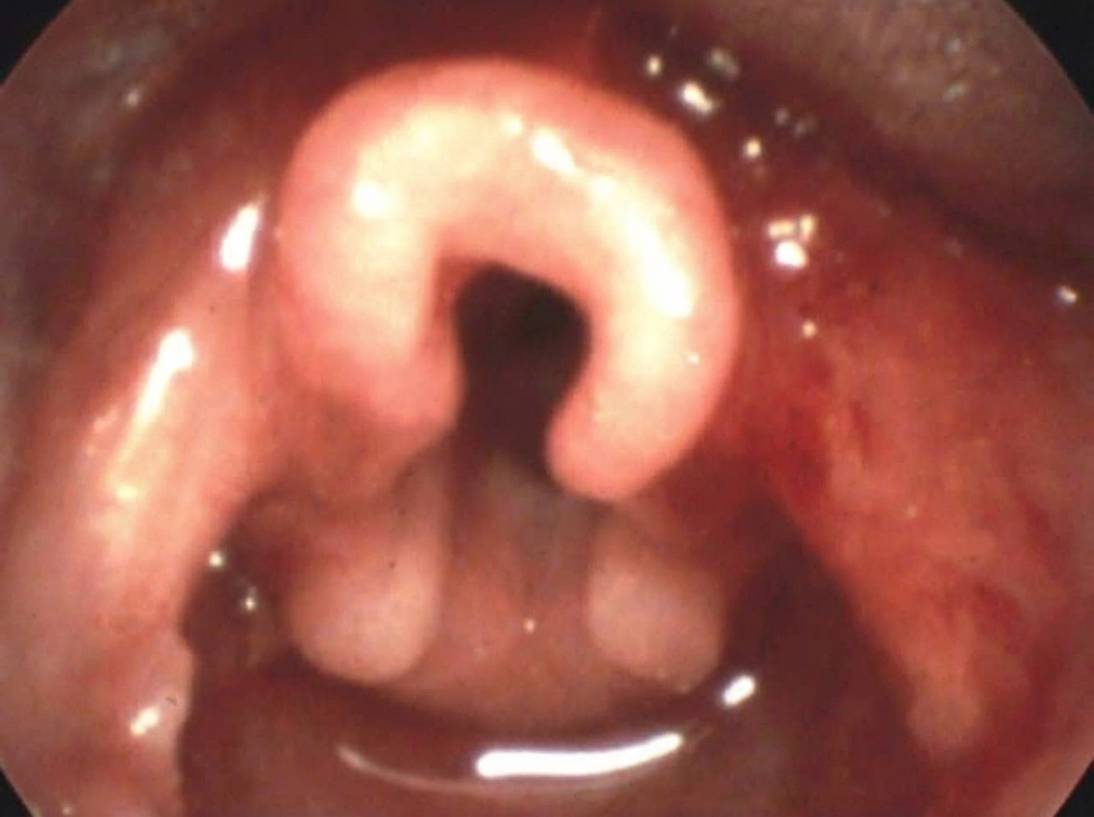
Which of the following is incorrect about the clinical condition shown below?

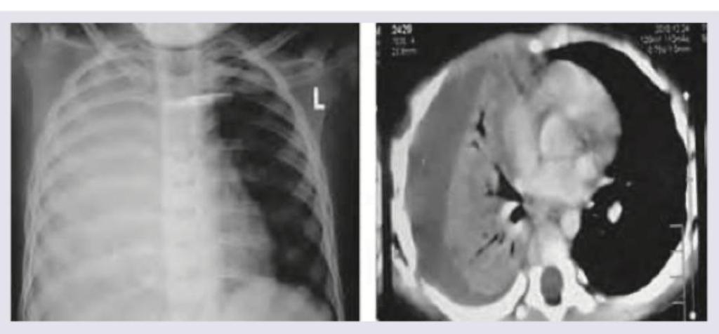
A 4-year-old child with high grade fever with chills and rigors for past 4 days and fast breathing is brought to the hospital. On examination subcostal recession and nasal flaring is noted. Since patient is already on antibiotics for past 3 days from another hospital a Chest X-ray and CT Chest was ordered. The diagnosis is:

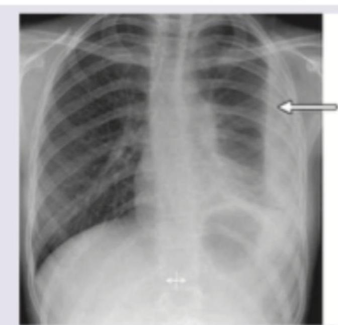
A 4-year-old admitted in ward with pneumonia. He develops sudden onset of breathlessness. What is the next step in management?

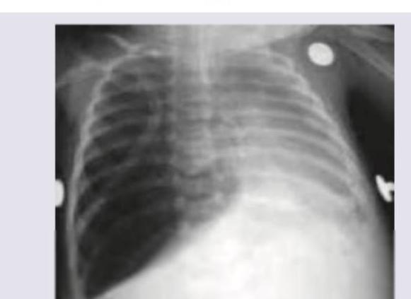
A 10-year-old child presents with fever, chills, rigors and fast, difficult breathing. The image shows:

A 12-month-old child who was previously well presented with low grade fever and respiratory distress and on examination there is bilateral wheeze. There is a history of URI in elder sibling. What is the diagnosis?
For a sick child aged 4 years, which of the following are signs of "severe pneumonia or very severe disease", as per IMNCI (Integrated Management of Neonatal and Childhood Illness) Program? I. Fast breathing (Greater than or equal to 40 breaths per minute) II. Child vomits everything III. Stridor in a calm child IV. Chest indrawing: Select the correct answer using the code given below :
A child of 2 years having a respiratory rate of 46 per minute shall be classified by a health worker as
Which sign is most important in deciding severe pneumonia in a child?
Explanation: ***Cystic fibrosis*** - This diagnosis is strongly supported by the triad of chronic **recurrent respiratory infections** (due to thick, retained sputum and subsequent bronchiectasis/bronchial wall thickening), and evidence of **pancreatic exocrine insufficiency** presenting as steatorrhea since birth. - It is an autosomal recessive disorder caused by mutations in the **CFTR gene**, leading to defective chloride transport and thick, sticky mucus formation in multiple organs. *Hyaline membrane disease* - Also known as **Respiratory Distress Syndrome (RDS)**, it is a condition primarily affecting **premature infants** due to deficiency of pulmonary surfactant. - It presents acutely within the first few hours of life and is not a cause of chronic, recurrent respiratory infections or steatorrhea in a 5-year-old child. *Alpha 1 anti-trypsin deficiency* - This hereditary condition typically presents with early-onset **panacinar emphysema** and/or liver disease (cirrhosis). - It is not associated with severe **pancreatic exocrine insufficiency** and steatorrhea in childhood, which is a hallmark feature of this patient's presentation. *Malabsorption syndrome* - While the patient exhibits findings consistent with **malabsorption** (steatorrhea), this is a general descriptive term and not a specific primary diagnosis for the entire clinical picture. - Cystic Fibrosis is the most specific primary diagnosis that explains both the **respiratory and gastrointestinal symptoms** (thick sputum, bronchial wall thickening, and steatorrhea).
Explanation: ***1, 3 and 4*** - This option correctly identifies the three most common and clinically significant causes of stridor in infants - **Laryngomalacia (1)** is the most common cause of congenital stridor (60-75% of cases), presenting with inspiratory stridor that worsens with agitation - **Subglottic stenosis (3)** is the second most common cause of congenital stridor, presenting with biphasic stridor and may be congenital or acquired - **Recurrent respiratory papillomatosis/RRP (4)** caused by HPV (types 6 and 11) causes progressive airway obstruction with stridor and hoarseness - While congenital (subglottic) hemangiomas can cause stridor, they are less common than the above three conditions and typically present between 1-3 months of age *Incorrect: 1 and 3* - This option is incomplete as it excludes **RRP (4)**, which is an important cause of pediatric airway obstruction - RRP typically presents in early childhood and requires serial surgical debulking *Incorrect: 2 and 3* - This option incorrectly excludes **Laryngomalacia (1)**, which is the single most common cause of stridor in infants - Missing laryngomalacia in the differential diagnosis would be a significant oversight *Incorrect: 1 and 4* - This option fails to include **Subglottic stenosis (3)**, the second most common cause of congenital stridor - Subglottic stenosis can be life-threatening and requires urgent evaluation and management
Explanation: ***Nocturnal cyanosis*** - **Nocturnal cyanosis is NOT a typical feature of laryngomalacia**, making this the correct answer to what is incorrect about the condition. Laryngomalacia is characterized by inspiratory stridor due to collapse of supraglottic structures, but it rarely causes cyanosis. - While symptoms may worsen during sleep, frank cyanosis indicates severe obstruction or hypoxemia, which is uncommon in typical laryngomalacia. If present, it suggests severe disease requiring surgical intervention. - Most cases of laryngomalacia are mild and self-limiting without significant oxygen desaturation. *Drooling of saliva* - Drooling is also not a typical feature of laryngomalacia, as the condition involves upper airway collapse, not difficulty with saliva management or swallowing. - However, **nocturnal cyanosis is the better answer** as it more clearly represents an atypical/severe presentation, whereas drooling is simply not associated with the condition at all. *Inspiratory stridor* - **Inspiratory stridor is the hallmark symptom** of laryngomalacia, caused by collapse of supraglottic structures during inspiration, leading to turbulent airflow. - This symptom is typically exacerbated by feeding, crying, agitation, or lying supine, and usually appears within the first 2 weeks to 2 months of life. *Omega-shaped epiglottis* - An **omega-shaped (Ω-shaped) epiglottis is a classic laryngoscopic finding** in laryngomalacia, visible during flexible laryngoscopy. - This anatomical configuration, along with redundant arytenoid mucosa and shortened aryepiglottic folds, contributes to the dynamic collapse during inspiration that causes stridor.
Explanation: ***Massive right sided parapneumonic effusion*** - The chest x-ray shows complete opacification of the right hemithorax with a **mediastinal shift to the left**, indicating a large volume of fluid in the right pleural space. - The CT scan confirms a large, homogeneous fluid collection on the right side, compressing the lung and mediastinum, consistent with a **massive pleural effusion**, likely parapneumonic given the clinical context of fever and respiratory distress despite antibiotics. *Massive pan consolidation* - While consolidation would appear opaque on X-ray, **massive pan consolidation** of an entire lung would typically not cause such a significant **mediastinal shift** as fluid does. - The CT images show a distinct **fluid-density collection** rather than diffuse parenchymal opacification characteristic of consolidation. *Right sided hydro-pneumothorax* - A hydro-pneumothorax would present with a **fluid level** on the chest X-ray and CT scan, indicating both air and fluid in the pleural space. - The images show a **homogeneous fluid collection** without any discernible air component or fluid level, ruling out pneumothorax. *Right sided segmental collapse* - Segmental collapse would typically involve only a portion of the lung and would manifest as a **volume loss** in a specific segment, often with features like crowding of vessels and bronchi. - The findings depict an entire hemithorax filled with fluid causing **volume expansion** on the affected side rather than collapse.
Explanation: ***Emergency needle thoracostomy*** - This patient, a 4-year-old with pneumonia and sudden breathlessness, likely has a **tension pneumothorax**, which is a life-threatening emergency requiring immediate decompression. The chest X-ray shows a collapsed right lung and a mediastinal shift, consistent with tension pneumothorax. - An **emergency needle thoracostomy** (needle decompression) is the immediate life-saving procedure to relieve the pressure in a tension pneumothorax before more definitive treatment can be initiated. - Performed by inserting a large-bore needle (14-16G) into the **2nd intercostal space, mid-clavicular line** on the affected side. *Intercostal drainage tube insertion* - While an intercostal drainage tube (chest tube) is the definitive treatment for pneumothorax, it takes more time to insert and is not the immediate first step for a **tension pneumothorax** in an unstable patient. - The delay in performing needle decompression could be fatal in a rapidly deteriorating patient with tension pneumothorax. *Decrease mechanical ventilation setting* - Decreasing mechanical ventilation settings would not address the underlying pathology of a tension pneumothorax, which is trapped air causing lung collapse and mediastinal shift. - This action could further compromise the patient's respiratory status if the pneumothorax is severe and the patient is already hypoxemic. *Increase mechanical ventilation setting* - Increasing mechanical ventilation settings would likely worsen a **tension pneumothorax** by forcing more air into the pleural space and increasing intrathoracic pressure. - This would further compromise venous return to the heart and reduce cardiac output, rapidly leading to **cardiovascular collapse**.
Explanation: ***Bacterial pneumonia with rupture of pneumatocele*** - The image shows a **large air-filled cystic lesion** (pneumatocele) in the left lung with evidence of surrounding **consolidation**, consistent with bacterial pneumonia. The arrow points to an area that suggests communication with the pleural space, indicative of rupture. - The clinical presentation of **fever, chills, rigors, and difficult breathing** in a child is highly suggestive of a severe bacterial lung infection, which can lead to complications such as pneumatoceles and their rupture. *Pleural effusion with collapse of lower lobe* - While there is some opacity on the left, the predominant feature is a **large air-filled cyst** rather than diffuse fluid collection. - A simple pleural effusion would typically show a **meniscus sign** and does not explain the large lucent area, which is characteristic of a pneumatocele or bulla. *Pneumonia with collapse* - Though there is evidence of pneumonia with consolidation, the key distinguishing feature here is the **large cystic structure (pneumatocele)**. - Lung collapse would present as significant **volume loss** and shifting of mediastinal structures, which is not the primary radiographic finding in this image. *Asthma* - Asthma is a **bronchospastic condition** and typically presents with hyperinflation on chest X-ray during an exacerbation, without discrete pulmonary lesions like pneumatoceles or consolidation. - The clinical symptoms of **fever, chills, and rigors** are not typical for uncomplicated asthma, but rather point towards an infectious etiology.
Explanation: ***Acute bronchiolitis*** - The clinical presentation of a **12-month-old** with **low-grade fever**, **respiratory distress**, and **bilateral wheeze**, along with the history of a preceding **URI in an elder sibling**, is highly characteristic of acute bronchiolitis, commonly caused by **Respiratory Syncytial Virus (RSV)**. - Bronchiolitis primarily affects infants and toddlers, causing inflammation and obstruction of the small airways, leading to wheezing and increased work of breathing. - The **age (12 months)** and **contact history** are key diagnostic clues. *Congestive cardiac failure* - While congestive cardiac failure can cause respiratory distress, it typically presents with other signs like **tachycardia**, **hepatomegaly**, and potentially a cardiac murmur, which are not mentioned here. - Wheezing can occur in cardiac failure but is usually accompanied by **rales** and signs of fluid overload rather than prominent diffuse wheezing as the primary respiratory finding in an otherwise well child. *Pneumonia* - Pneumonia would more typically present with a **higher fever**, **cough**, and focal lung findings such as **crackles** or **dullness to percussion**, rather than predominantly bilateral wheezing. - Chest X-ray in pneumonia often shows **infiltrates** or **consolidation**, whereas in bronchiolitis, it commonly shows hyperinflation or peribronchial thickening. *Asthma exacerbation* - Asthma is uncommon as a first diagnosis in a child under **2 years of age**, especially in a **previously well** child with no history of recurrent wheezing episodes. - The **contact history with URI in a sibling** and the **acute, first-time presentation** strongly favor an infectious etiology (bronchiolitis) rather than asthma. - Asthma exacerbations typically occur in children with a known history of reactive airway disease and recurrent wheezing episodes.
Explanation: ***III and IV*** - **Stridor in a calm child** is a critical sign indicating severe upper airway obstruction, classified as "very severe disease" in IMNCI. This is a general danger sign requiring urgent referral. - **Chest indrawing** (severe lower chest wall indrawing) signifies increased work of breathing and severe respiratory distress, categorizing it under "severe pneumonia" in IMNCI and requiring urgent referral. - These two signs represent the most specific respiratory indicators of severe pneumonia or very severe disease. *I and II* - **Fast breathing** for a 4-year-old (age 12 months to 5 years) is correctly defined as ≥40 breaths per minute per IMNCI. However, fast breathing alone indicates **pneumonia** (not severe pneumonia), which can be managed as outpatient with oral antibiotics. - **Child vomits everything** is indeed a general danger sign indicating "very severe disease" in IMNCI, but when combined with fast breathing (which indicates only simple pneumonia), this combination is less specific than III and IV. *II and III* - **Child vomits everything** is a general danger sign for "very severe disease" requiring urgent referral. - **Stridor in a calm child** is also a sign of "very severe disease." - While both are valid signs, this combination misses **chest indrawing**, which is the primary respiratory-specific sign of severe pneumonia and more directly answers the question about severe pneumonia classification. *II and IV* - **Child vomits everything** is a general danger sign for "very severe disease." - **Chest indrawing** is the key sign of "severe pneumonia." - While both are valid signs of severe pneumonia or very severe disease, the combination of **stridor and chest indrawing (III and IV)** represents the two most specific respiratory signs and is the preferred answer for this classification.
Explanation: ***Correct: Pneumonia*** - A respiratory rate of **46 breaths per minute** in a 2-year-old child falls within the criteria for **fast breathing**. According to World Health Organization (WHO) IMCI guidelines, fast breathing is defined as a respiratory rate ≥ 50 breaths/minute for children aged 2 months to 12 months, and **≥ 40 breaths/minute for children aged 12 months to 5 years**. - Fast breathing alone (without chest indrawing or danger signs) is the **key clinical sign** for classifying a child with cough or difficulty breathing as having **pneumonia**. - This is based on the **WHO IMCI classification** used by health workers for management of childhood illness. *Incorrect: No pneumonia, cough or cold* - This classification would be made if the child's respiratory rate was **within the normal range** for their age (< 40 breaths per minute for age 1-5 years) and if there were no other signs of pneumonia or severe disease. - A respiratory rate of 46 breaths per minute in a 2-year-old is **above the normal limit** and meets the criteria for fast breathing. *Incorrect: Severe pneumonia* - Severe pneumonia is classified by the presence of **chest indrawing** in addition to cough or difficult breathing, without danger signs. - While the child has fast breathing, the question does not mention **chest indrawing**, which is required for this classification. *Incorrect: Very severe disease* - Very severe disease is classified when there are **danger signs** present: inability to drink or breastfeed, persistent vomiting, convulsions, lethargy or unconsciousness, or stridor in a calm child. - The question only mentions elevated respiratory rate without any **danger signs**, so this classification does not apply.
Explanation: ***Chest indrawing*** - **Chest indrawing** (or retractions) indicates increased work of breathing due to reduced lung compliance or airway obstruction, a key sign of severe pneumonia in children. - It signifies that accessory muscles are engaged to pull the chest wall inward with each breath, demonstrating significant respiratory distress. *Fast breathing* - **Fast breathing** (tachypnea) is a general sign of respiratory distress and a criterion for diagnosing pneumonia, but it alone does not differentiate severe from non-severe pneumonia. - While present in pneumonia, it doesn't indicate the same level of respiratory effort or severity as chest indrawing. *Nasal flaring* - **Nasal flaring** is an effort to decrease airway resistance by widening the nostrils during inspiration, indicating increased work of breathing. - It's a sign of respiratory distress that can occur in both moderate and severe pneumonia, but it is not as specific to severe disease as chest indrawing. *Grunting* - **Grunting** is an expiratory sound produced by partial closure of the glottis to maintain positive end-expiratory pressure, preventing alveolar collapse. - While a sign of moderate to severe respiratory distress, especially in neonates, it does not carry the same weight as chest indrawing for classifying severe pneumonia in older children by WHO guidelines.
Upper Respiratory Tract Infections
Practice Questions
Lower Respiratory Tract Infections
Practice Questions
Asthma Management
Practice Questions
Cystic Fibrosis
Practice Questions
Bronchiolitis
Practice Questions
Foreign Body Aspiration
Practice Questions
Sleep-Disordered Breathing
Practice Questions
Congenital Lung Malformations
Practice Questions
Pleural Diseases
Practice Questions
Tuberculosis in Children
Practice Questions
Chronic Lung Disease in Premature Infants
Practice Questions
Pulmonary Function Testing
Practice Questions
Get full access to all questions, explanations, and performance tracking.
Start For Free