
Pediatric Surgery Basics — MCQs

On this page
In congenital diaphragmatic hernia, which of the following is not typically seen?
A 6-week-old boy presents with a palpable abdominal mass in the epigastrium. The clinical diagnosis is:
A 3-week-old child presents with projectile postprandial vomiting and is diagnosed with hypertrophic pyloric stenosis. Congenital hypertrophic pyloric stenosis is associated with which of the following acid-base and electrolyte disturbances?
What is true about cystic hygroma?
Gasless abdomen on X-ray is seen with what type of tracheo-esophageal fistula?
A child is born with bilious vomiting and failure to thrive. Which of the following is true about this condition?
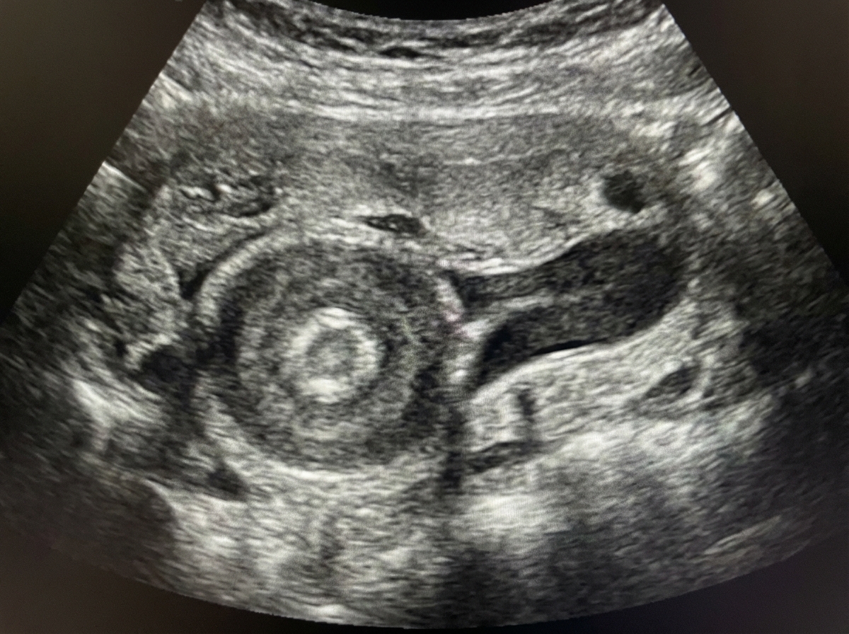
Prenatal ultrasound at 20 weeks revealed a midline mass that appeared to contain intestines and was membrane-bound. What is the probable diagnosis?
What is the probable diagnosis for a cyst in a child that is located at and associated with vertebral defects?
In a 2-year-old child, what is the most likely diagnosis for a bluish, dome-shaped swelling on the inner side of the lip?
Which of the following is false regarding Bochdalek hernia?
Practice by Chapter
Surgical Conditions of the Newborn
Practice Questions
Congenital Diaphragmatic Hernia
Practice Questions
Esophageal Atresia and Tracheoesophageal Fistula
Practice Questions
Intestinal Atresia and Stenosis
Practice Questions
Malrotation and Volvulus
Practice Questions
Hirschsprung's Disease
Practice Questions
Anorectal Malformations
Practice Questions
Biliary Atresia
Practice Questions
Abdominal Wall Defects
Practice Questions
Inguinal Hernia and Hydrocele
Practice Questions
Intussusception
Practice Questions
Appendicitis in Children
Practice Questions
Want unlimited practice?
Get full access to all questions, explanations, and performance tracking.
Scan to download app