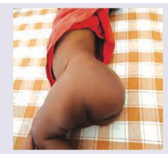
A 2-month-old girl was brought with swelling just above the gluteal area with progressive increase in size. Which is the most probable diagnosis?

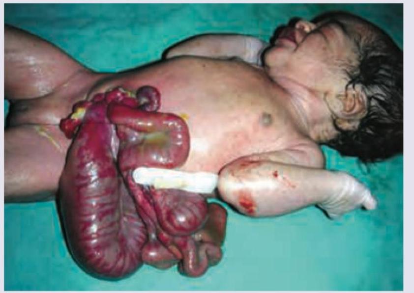
The image shows presence of:

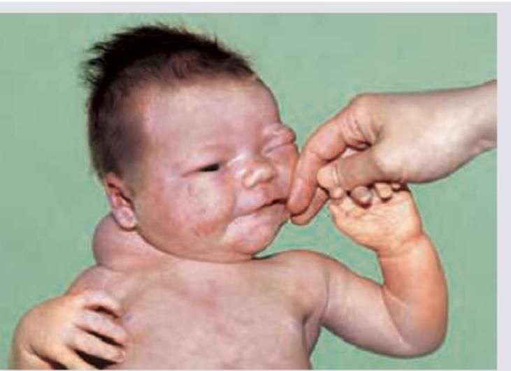
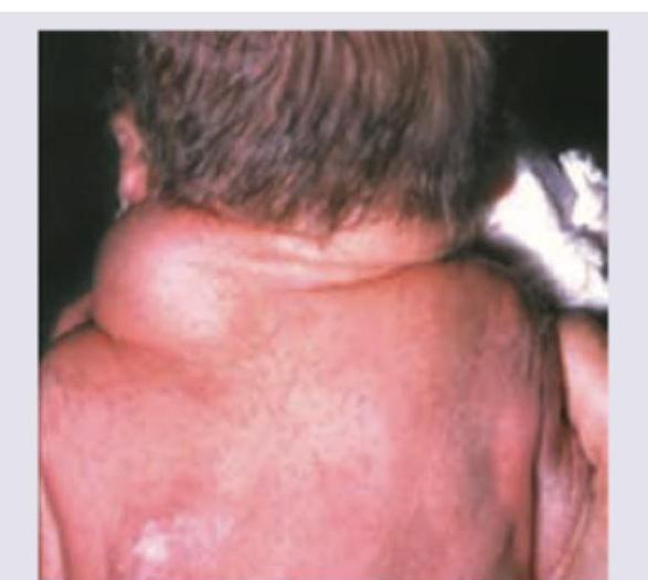
A neonate presents with a soft, compressible, translucent cystic mass in the neck region, present since birth. What is the most likely diagnosis?

A 3-year-old child presents with swelling in scrotum since birth. Transillumination test is positive. All are true except:
Which of the following is the first line of management of the condition shown?

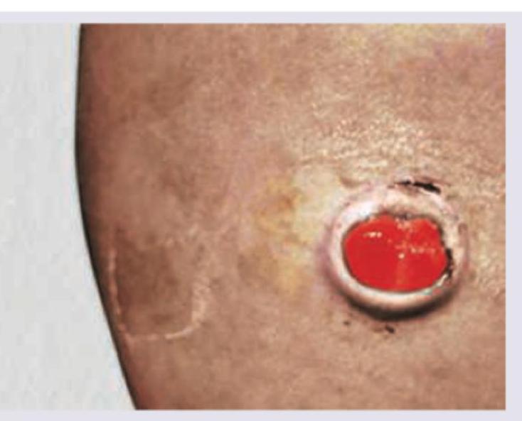
Which of the following is incorrect about the image shown?

What does the following image show?

A 5-year-old male child comes with a left sided scrotal swelling which has no cough impulse and does not reduce on compression or lying down but the parents give a definite history that swelling is absent in the morning and comes by in the evening. The best treatment is :
Most common cause of intestinal obstruction in childhood is:
Consider the following statements in respect of congenital hypertrophic pyloric stenosis: 1. The condition is more common in males. 2. The investigation of choice is ultrasonography. 3. Hypertrophy is maximal in the pre pyloric region. 4. Projectile vomiting is seen in this condition. Which of the statements given above are correct?
Explanation: ***Sacrococcygeal teratoma*** - Sacrococcygeal teratomas are the **most common germ cell tumors in neonates and infants**, often presenting as a mass in the sacral region, as seen in the image. - They arise from pluripotent cells in the **Hensen's node** (primitive streak remnant) and can contain tissues derived from all three germ layers. *Rhabdomyosarcoma* - Rhabdomyosarcoma is a **malignant tumor of skeletal muscle origin** that can occur in various locations, including the gluteal area, but is less common in infants than SCT. - While it can present as a rapidly growing mass, a sacrococcygeal location in an infant points more strongly to teratoma. *Atypical Rhabdoid tumor* - Atypical rhabdoid tumor is a **highly malignant tumor** typically found in the brain and kidney, though it can occur in soft tissues. - It is rare and not typically the primary diagnosis for a large external gluteal mass in a 2-month-old. *Fibrosarcoma* - Fibrosarcoma is a **malignant tumor of fibroblasts**, rare in infants and usually occurring in older children or adults. - It would present as a soft tissue mass but is much less common than a sacrococcygeal teratoma in this age group and location.
Explanation: ***Gastroschisis*** - The image shows **exposed abdominal organs** (intestines) protruding through a defect in the abdominal wall, typically to the **right of the umbilical cord**. - In gastroschisis, there is **no covering membrane** over the protruding organs, which is characteristic of the condition seen in the image. *Omphalocele* - An omphalocele involves the **protrusion of abdominal organs** through the umbilical cord, but they are contained within a **peritoneal-amniotic sac**. - The image clearly shows **uncovered intestines**, ruling out an omphalocele. *Necrotizing enterocolitis* - This condition is an **inflammatory bowel disease** affecting premature infants, characterized by bowel necrosis, not by external protrusion of organs. - Clinical signs would include abdominal distension, feeding intolerance, and bloody stools, which are not directly depicted or implied here. *Malformation of vitello-intestinal duct* - Malformations of the vitello-intestinal duct (e.g., Meckel's diverticulum or persistent vitelline duct) involve remnants of the embryonic yolk sac connection to the intestine. - These malformations do not typically present as a large external defect with exposed bowel loops as seen in the image.
Explanation: ***Lymphangioma*** - The clinical presentation of a **soft, compressible, translucent cystic mass** in the neck of a neonate is characteristic of **cervical lymphangioma** (also known as cystic hygroma) - These are **congenital malformations of the lymphatic system** that typically present at birth or within the first two years of life - The mass is usually **transilluminable** due to its fluid-filled nature, which is a key diagnostic feature - Most commonly located in the **posterior triangle of the neck** *Branchial cyst* - **Branchial cleft cysts** present as lateral neck masses, typically anterior to the sternocleidomastoid muscle - They usually become **clinically apparent later in childhood** or adolescence, often after an upper respiratory infection - The mass is more **circumscribed and firm**, not typically transilluminant like lymphangiomas - Less common in neonates compared to lymphangiomas *Hemangioma* - **Infantile hemangiomas** can present as neck masses but typically have a **vascular appearance** with overlying skin changes (red or purple discoloration) - They are **not transilluminant** and feel more solid or rubbery rather than cystic - Hemangiomas often show **rapid growth phase** during early infancy (proliferative phase) followed by involution - They do not typically present as translucent cystic masses *Mumps* - **Mumps** causes painful swelling of the **parotid glands**, located in front of and below the ears - It is a **viral infection** (paramyxovirus), not a congenital condition - Presentation in neonates is **extremely rare** due to maternal antibody protection - The swelling is **bilateral**, tender, and associated with systemic symptoms (fever, malaise), not a solitary translucent mass
Explanation: ***Bag of worms*** - A "bag of worms" sensation on palpation of the scrotum is characteristic of a **varicocele**, which is an enlargement of the pampiniform plexus of testicular veins. - The presented case describes a **hydrocele**, characterized by scrotal swelling since birth and positive transillumination, which is a collection of fluid, not dilated veins. *Herniotomy* - A persistent **patent processus vaginalis** is the underlying cause of a communicating infantile hydrocele. - **Herniotomy** is the surgical procedure performed to ligate and excise the patent processus vaginalis, thereby treating the hydrocele and preventing future herniation. *USG for patent processus vaginalis* - **Ultrasound (USG)** is a common diagnostic tool used to assess scrotal swelling and can identify a patent processus vaginalis, which is the communication between the peritoneal cavity and the tunica vaginalis, responsible for a communicating hydrocele. - This imaging confirms the presence of fluid within the tunica vaginalis and, potentially, the communicating channel, helping to differentiate from other causes of scrotal swelling. *Does not reduce on supine position* - A typical **communicating hydrocele** may fluctuate in size but generally **does not reduce** completely on a change in position, unlike an inguinal hernia which often reduces when the child is supine. - This is because the fluid within the tunica vaginalis cannot easily drain back into the peritoneal cavity, especially if the communication is small or intermittently obstructed.
Explanation: ***Parenteral vitamin K*** - The image shows an actively **bleeding umbilical stump**, which is a classic presentation of **vitamin K deficiency bleeding** (VKDB) in a neonate. - **Parenteral vitamin K** is the first-line treatment for VKDB because it rapidly replenishes clotting factors, addressing the underlying coagulopathy. *Silver nitrate topical application* - **Silver nitrate** is used to cauterize and treat **umbilical granulomas**, which appear as pinkish-red, moist, fleshy growths, typically not actively bleeding. - It would be ineffective and potentially harmful for an actively bleeding umbilical stump due to vitamin K deficiency. *Ligation of stump with silk thread* - **Ligation with a silk thread** is indicated for a persistent, non-bleeding **umbilical granuloma** that is not responsive to silver nitrate. - It is not appropriate for treating active bleeding due to coagulopathy and could worsen the condition if the underlying cause is not addressed. *Topical neosporin powder* - **Topical neosporin powder** is an antibiotic used for preventing **bacterial infections** of the umbilical stump or minor skin wounds. - It does not have any hemostatic properties and would not stop bleeding caused by a systemic clotting deficiency.
Explanation: ***Always presents with stridor*** - This statement is **incorrect** because stridor is **not a universal presentation** of cystic hygroma. - Stridor only develops when the lesion is large enough or positioned to compress the airway (typically **cervical location near trachea**). - Many cystic hygromas, particularly **smaller ones** or those located away from respiratory structures, do **not present with stridor** at all. - Other presentations include **painless neck mass**, **dysphagia**, or **incidental prenatal finding**. *Occurs due to failure of lymphatics to connect to venous system* - This accurately describes the **primary embryological defect** in cystic hygroma formation. - Primitive lymphatic sacs fail to connect to the venous system, leading to **accumulation of lymphatic fluid** and formation of the characteristic multiloculated cystic mass. *Associated with Turner syndrome* - Cystic hygromas are strongly associated with **Turner syndrome (45,X)**, particularly **nuchal cystic hygromas** detected prenatally. - Other chromosomal abnormalities such as **trisomy 13, 18, and 21** also show increased association with cystic hygromas. *CT scan for mediastinal extension* - **CT or MRI imaging** is essential for assessing the full extent of cystic hygromas and identifying **mediastinal, laryngeal, or airway involvement**. - This imaging is crucial for **surgical planning** and anticipating potential complications such as respiratory compromise.
Explanation: ***Invertogram*** - An invertogram is a **lateral radiograph** of the abdomen with the patient **held upside down** to visualize the distal bowel gas pattern and evaluate for imperforate anus. - The image shows dilated bowel loops with gas, and the child's position (evident from the spinal curvature) is consistent with an inverted view. *Infantogram* - This term is **not a standard medical diagnostic procedure** or specific type of radiograph. - While it refers to an X-ray of an infant, it does not describe the specific technique or purpose shown. *Gynogram* - A gynogram is an older term for a radiological procedure involving the **injection of contrast material** to visualize female pelvic organs, particularly the uterus and fallopian tubes. - It is unrelated to the gastrointestinal tract or the patient's position in this image. *Vasogram* - A vasogram, also known as **vasography**, is a procedure that involves injecting contrast into the **vas deferens** or other vascular structures to visualize them. - This technique is used to assess male reproductive structures or blood vessels and is not applicable to evaluating bowel obstruction in an infant as depicted.
Explanation: ***Herniotomy*** - This presentation is classic for a **communicating hydrocele** in a 5-year-old child, where peritoneal fluid accumulates in the scrotum through a **patent processus vaginalis** during the day (when upright) and drains back into the peritoneal cavity overnight (when recumbent), explaining the absence in morning and presence in evening. - The absence of cough impulse and lack of reducibility on compression distinguishes this from a typical inguinal hernia, but the fluctuating size pattern confirms communication with the peritoneal cavity. - **Management:** While communicating hydroceles may resolve spontaneously in infancy (typically by 12-18 months), **persistence beyond 2 years of age is an indication for surgical repair**. At **age 5**, surgical correction is clearly indicated. - **Herniotomy** with **high ligation of the patent processus vaginalis** is the treatment of choice. This procedure closes the communication between the peritoneal cavity and the tunica vaginalis, preventing further fluid accumulation. - The processus vaginalis is ligated at the internal inguinal ring, and the distal sac is left open to allow reabsorption of any residual fluid. *To leave it alone (masterly inactivity)* - **Observation (masterly inactivity)** is appropriate for communicating hydroceles in **infants under 12-18 months** as spontaneous closure of the processus vaginalis commonly occurs. - However, at **age 5 years**, the likelihood of spontaneous resolution is extremely low, and continued observation would be inappropriate. - Persistent patent processus vaginalis carries a risk of developing an indirect inguinal hernia, making surgical intervention the standard of care at this age. *Eversion of sac* - **Eversion of the sac** (Jaboulay's procedure) or plication (Lord's procedure) is used for **non-communicating hydroceles in adults** where the processus vaginalis is obliterated. - These procedures are **not appropriate for communicating hydroceles in children** as they do not address the underlying patent processus vaginalis. - Without ligating the patent communication, fluid will continue to accumulate from the peritoneal cavity. *Hernioplasty* - **Hernioplasty** typically refers to hernia repair with **mesh reinforcement**, which is an adult procedure. - In pediatric inguinal region surgery, mesh is generally **avoided** due to concerns about growth, tissue reaction, and long-term complications. - The pediatric approach focuses on simple high ligation of the sac (herniotomy) rather than mesh repair (hernioplasty).
Explanation: ***Intussusception*** - **Intussusception** is the most common cause of intestinal obstruction in children between **3 months and 6 years** of age, particularly in infants. - It involves the telescoping of one segment of the intestine into an adjacent segment, leading to obstruction and potentially **ischemia**. *Mucoviscidosis* - **Mucoviscidosis**, or **cystic fibrosis**, can cause intestinal obstruction in newborns due to **meconium ileus**, but it is less common across the entire childhood period than intussusception. - While it is a significant cause of intestinal issues, its prevalence as the *most common overall cause* of obstruction in childhood is lower. *Hirschsprung's disease* - **Hirschsprung's disease** is a congenital absence of **ganglion cells** in the distal bowel, leading to functional obstruction, primarily in **neonates** and infants. - While an important cause of obstruction in early life, its overall incidence is less than intussusception, especially when considering the broader childhood age range. *Volvulus* - **Volvulus** is the twisting of a section of the intestine around its mesentery, which can occur at any age but is less frequent than intussusception in childhood. - It often presents as an acute surgical emergency with severe symptoms, but it is not the *most common* cause of obstruction across childhood.
Explanation: ***1, 2 and 4 only*** - **Statement 1 is CORRECT**: Congenital hypertrophic pyloric stenosis (CHPS) is more common in males with a male-to-female ratio of approximately 4-5:1. - **Statement 2 is CORRECT**: Ultrasonography is the investigation of choice for CHPS, being non-invasive and accurate in measuring pyloric muscle thickness (>3 mm) and pyloric length (>15 mm). - **Statement 3 is INCORRECT**: The hypertrophy is maximal in the **pyloric muscle** (circular muscle layer of the pylorus), NOT in the pre-pyloric region. This is a key anatomical distinction. - **Statement 4 is CORRECT**: Projectile non-bilious vomiting typically occurring 30-60 minutes after feeding is the hallmark clinical presentation of CHPS. *3 only* - Incorrect because statement 3 is false (hypertrophy is in the pylorus, not pre-pyloric region), while statements 1, 2, and 4 are all true. *2, 3 and 4 only* - Incorrect because it includes statement 3, which is false. The maximal hypertrophy occurs in the **pyloric canal**, not the pre-pyloric region. *1 and 2 only* - Incomplete as it omits statement 4 about projectile vomiting, which is a cardinal feature of CHPS and is definitely correct.
Surgical Conditions of the Newborn
Practice Questions
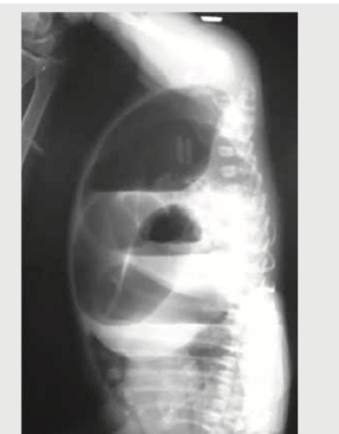
Congenital Diaphragmatic Hernia
Practice Questions
Esophageal Atresia and Tracheoesophageal Fistula
Practice Questions
Intestinal Atresia and Stenosis
Practice Questions
Malrotation and Volvulus
Practice Questions
Hirschsprung's Disease
Practice Questions
Anorectal Malformations
Practice Questions
Biliary Atresia
Practice Questions
Abdominal Wall Defects
Practice Questions
Inguinal Hernia and Hydrocele
Practice Questions
Intussusception
Practice Questions
Appendicitis in Children
Practice Questions
Get full access to all questions, explanations, and performance tracking.
Start For Free