What is the most common type of congenital atresia?
Which electrolyte should be replenished in hypertrophic pyloric stenosis?
What is true about Bochdalek hernia?
A 9-month-old girl presents with perianal bleeding, vomiting, a right lumbar mass, and masked liver dullness. She is in a shock-like condition. Management should include all of the following EXCEPT?
Which of the following is true for Bochdalek hernia?
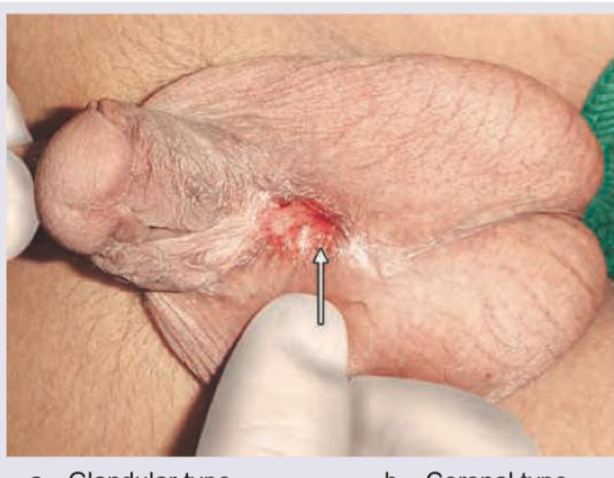
The image shows:

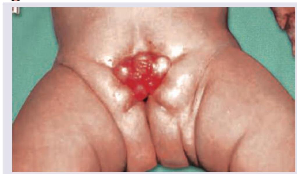
What is the malformation shown in the image?

The image shows:

A newborn presents with respiratory distress shortly after birth. The chest X-ray shows bowel loops in the thoracic cavity and absence of bowel loops in the abdominal area. There is associated hypoplasia of the ipsilateral lung. What is the most likely diagnosis?

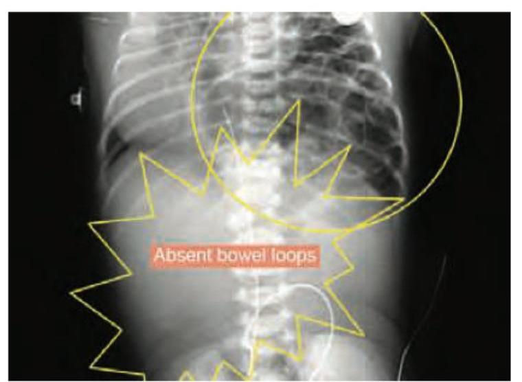
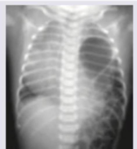
All are true about the condition shown in the figure except:

Explanation: This question refers to **Tracheoesophageal Fistula (TEF) and Esophageal Atresia (EA)**, a high-yield topic in pediatric surgery. ### **Explanation of the Correct Answer** The most common variant is **Type C** (Vogt-Ladder classification) or **Type IIIb** (Gross classification). It accounts for approximately **85% of all cases**. In this type, the upper esophagus ends in a **blind pouch** (Atresia), while the lower esophagus communicates with the trachea via a **fistula** (distal TEF). **Pathophysiology:** During the 4th–5th week of gestation, the tracheoesophageal septum fails to divide the foregut completely. In Type C, the distal fistula allows air to enter the stomach (causing abdominal distension) and gastric acid to reflux into the lungs (causing aspiration pneumonia). ### **Analysis of Incorrect Options** * **Option B (Type B):** This describes a proximal fistula with a distal blind end. It is rare (~1%) and often presents with early, severe aspiration during feeding. * **Option C (Type E/H-type):** This is an "H-type" fistula where the esophagus is continuous (no atresia) but connected to the trachea. It presents later in childhood with recurrent pneumonia and "cyanotic spells" while feeding. * **Option D:** This is clinically irrelevant and does not describe a recognized anatomical variant of TEF/EA. ### **Clinical Pearls for NEET-PG** * **Most common type:** Type C (Proximal Atresia + Distal Fistula). * **Pure Atresia (Type A):** Second most common (~8%); characterized by a **gasless abdomen** on X-ray. * **Clinical Presentation:** Drooling of saliva, choking/cyanosis on first feed, and inability to pass a nasogastric tube (coils in the pouch). * **Associated Anomalies:** **VACTERL** association (Vertebral, Anal, Cardiac, TEF, Renal, Limb) is present in 50% of cases. * **Investigation of Choice:** Plain X-ray (Chest + Abdomen) with a radio-opaque catheter in situ.
Explanation: ### Explanation **Correct Answer: C. Chloride** In **Infantile Hypertrophic Pyloric Stenosis (IHPS)**, the hallmark metabolic derangement is **Hypochloremic, Hypokalemic, Metabolic Alkalosis with Paradoxical Aciduria**. The primary event is persistent non-bilious projectile vomiting, which leads to a massive loss of gastric secretions. These secretions are rich in **Hydrochloric acid (HCl)** and water. The loss of Chloride ($Cl^-$) is the most significant electrolyte deficit. As chloride levels drop, the kidneys attempt to maintain electrical neutrality by reabsorbing Bicarbonate ($HCO_3^-$), which worsens the alkalosis. Therefore, replenishing **Chloride** (usually via Normal Saline) is the most critical step in correcting the metabolic state before surgery. **Analysis of Incorrect Options:** * **A. Sodium:** While sodium is lost in the vomitus, the body’s primary deficit that drives the complex metabolic alkalosis is Chloride. Sodium is replaced as the carrier (Normal Saline), but Chloride is the specific ion required to break the cycle of bicarbonate retention. * **B. Hydrogen:** Hydrogen ions are lost in the vomitus (causing alkalosis). We do not "replenish" hydrogen; instead, we provide Chloride, which allows the kidneys to excrete excess bicarbonate, thereby restoring the pH. * **D. Bicarbonate:** Bicarbonate levels are already **elevated** (Metabolic Alkalosis). Adding more would be dangerous and exacerbate the condition. **High-Yield Clinical Pearls for NEET-PG:** * **The "Paradoxical Aciduria":** In late stages, the body prioritizes volume over pH. To save $Na^+$, the kidney exchanges it for $H^+$ in the distal tubule (due to depleted $K^+$), leading to acidic urine despite systemic alkalosis. * **Diagnosis:** Best initial investigation is **Ultrasonography** (Target sign/Donut sign). * **Physical Exam:** Palpable **"Olive-shaped mass"** in the epigastrium. * **Surgery:** Ramstedt’s Pyloromyotomy (Note: This is a medical emergency, not a surgical one; correct electrolytes first!).
Explanation: **Explanation:** **Bochdalek Hernia** is the most common type of congenital diaphragmatic hernia (CDH), resulting from the failure of the pleuroperitoneal canal to close during embryonic development. **Why Option C is correct:** A Bochdalek hernia typically presents as a mass in the posterior mediastinum or the cardiophrenic angle. On imaging (like a chest X-ray or CT), a fat-containing hernia or a hernia involving abdominal viscera can mimic other cystic or solid lesions in the lower mediastinum. Therefore, it is a recognized **differential diagnosis for a pleuropericardial cyst**, as both can appear as masses near the diaphragm/heart borders. **Why other options are incorrect:** * **Option A:** Bochdalek hernias are overwhelmingly **left-sided (approx. 80-85%)**. The right side is protected by the presence of the liver, which acts as a physical barrier during development. * **Option B:** It is located **posterolaterally**. In contrast, the **Morgagni hernia** is the one located in an anterior (retrosternal) position. **High-Yield Clinical Pearls for NEET-PG:** * **Mnemonic:** **B**ochdalek is **B**ack and **B**ig (Posterior and more common); **M**orgagni is **M**idline/Anterior and **M**ore rare. * **Clinical Triad:** Scaphoid abdomen, respiratory distress, and shifted heart sounds (dextrocardia if left-sided). * **Most Important Prognostic Factor:** The degree of **pulmonary hypoplasia** and associated pulmonary hypertension, rather than the defect size itself. * **Management:** It is a physiological emergency, not a surgical one. Stabilize the infant (intubation, avoid bag-mask ventilation) before considering surgical repair.
Explanation: **Explanation:** The clinical presentation of a 9-month-old with perianal bleeding (likely "red currant jelly" stools), vomiting, and a palpable right lumbar mass is classic for **Intussusception**. However, the presence of **masked liver dullness** and a **shock-like condition** indicates a critical complication: **bowel perforation and peritonitis.** **Why Barium Enema is the Correct Answer (Contraindicated):** In stable cases of intussusception, a barium or air enema is the gold standard for both diagnosis and non-surgical reduction. However, if there are signs of **peritonitis, perforation (pneumoperitoneum causing masked liver dullness), or profound shock**, any form of hydrostatic or pneumatic reduction is **strictly contraindicated**. Performing an enema in this state increases intra-luminal pressure, which can worsen the perforation and lead to tension pneumoperitoneum or fecal contamination of the peritoneal cavity. **Why the other options are incorrect (Necessary Management):** * **Intravenous fluids:** Essential for resuscitation to manage the shock-like state and correct dehydration/electrolyte imbalances. * **Oxygen administration:** Necessary to ensure adequate tissue perfusion in a patient in shock. * **Nasogastric tube insertion:** Crucial for gastric decompression to prevent aspiration and reduce abdominal distension prior to surgery. **NEET-PG High-Yield Pearls:** * **Triad of Intussusception:** Paroxysmal pain, sausage-shaped mass (usually right upper quadrant/lumbar), and red currant jelly stools. * **Dance’s Sign:** An "empty" right iliac fossa due to the migration of the cecum. * **Target/Donut Sign:** The classic appearance on ultrasonography (the investigation of choice). * **Management Rule:** If the patient is stable, attempt non-surgical reduction (Air/Hydrostatic). If the patient has peritonitis or shock, proceed directly to **emergency laparotomy**.
Explanation: **Explanation:** **Bochdalek Hernia** is the most common type of Congenital Diaphragmatic Hernia (CDH), accounting for approximately 85-90% of cases. 1. **Why Option B is Correct:** It is a **congenital disorder** resulting from the failure of the pleuroperitoneal membranes to fuse with the septum transversum and dorsal mesentery of the esophagus during the 8th to 10th week of intrauterine life. This creates a defect in the diaphragm through which abdominal viscera herniate into the thoracic cavity. 2. **Why Other Options are Incorrect:** * **Option A:** It occurs most commonly on the **left side (80-85%)**. The right side is protected by the presence of the liver. * **Option C:** It is a **posterolateral** defect. Anterior defects are known as **Morgagni hernias**, which are much rarer. * **Option D:** It typically causes **immediate respiratory distress** (within minutes to hours of birth), not starting on day 2-3. The distress is due to pulmonary hypoplasia and persistent pulmonary hypertension. **High-Yield Clinical Pearls for NEET-PG:** * **Triad of CDH:** Dyspnea, Cyanosis, and Dextrocardia (if left-sided). * **Physical Exam:** Scaphoid abdomen, absent breath sounds on the affected side, and bowel sounds heard in the chest. * **Radiology:** "Bowel loops in the hemithorax" with a mediastinal shift. * **Management:** Initial stabilization is key. **Avoid bag-and-mask ventilation** (distends the gut, worsening lung compression); instead, perform immediate endotracheal intubation. Surgery is not an emergency; it is performed after the neonate is hemodynamically stable.
Explanation: ***Hesitation cuts*** - The image displays multiple, superficial, parallel incised wounds, primarily on the wrist, which are classic characteristics of **hesitation cuts**. - These wounds are often made by individuals contemplating **suicide** and are typically not immediately life-threatening, reflecting a tentative or experimental attempt before a deeper, fatal cut. *Defense wounds* - **Defense wounds** typically appear on the hands, forearms, and sometimes the legs, resulting from an individual instinctively shielding themselves from an assault. - They tend to be more varied in depth and pattern, often showing signs of struggle and resistance, unlike the uniform, superficial nature seen here. *Radiation injury* - **Radiation injury** or radiation dermatitis presents with skin changes ranging from erythema, desquamation, blistering, or hyperpigmentation, depending on the dose and duration of exposure. - These injuries do not typically involve sharp, incised wounds like those depicted in the image. *Homicidal stab* - A **homicidal stab** implies a deep, forceful wound intended to cause death, often characterized by a single or few much deeper penetrating wounds. - The superficial and numerous nature of the cuts in the image is inconsistent with the typical presentation of a homicidal attack.
Explanation: ***Penoscrotal type*** - The image distinctly shows the **urethral opening** located on the **ventral aspect of the penis**, at the junction where the penis meets the scrotum. - This position, where the meatus is located proximally at the base of the penis or in the scrotum, is characteristic of the **penoscrotal type of hypospadias**. *Glandular type* - In **glandular hypospadias**, the urethral opening is located on the **glans penis**, typically slightly displaced from the tip. - The image shows the opening much further down the shaft, not on the glans. *Coronal type* - The **coronal type of hypospadias** refers to the urethral opening being located in the **corona**, the groove separating the glans from the shaft of the penis. - The opening in the image is more proximal than the coronal sulcus. *Penile type* - **Penile hypospadias** refers to the urethral opening being located anywhere along the **shaft of the penis**, but it is a broad category. - The specific location shown in the image, at the base of the penis near the scrotum, is more precisely described as **penoscrotal hypospadias**.
Explanation: ***Bladder exstrophy*** - The image clearly displays an **exposed bladder mucosa** projecting from the lower abdominal wall, which is characteristic of bladder exstrophy. - This congenital anomaly results from a **failure of fusion of the anterior abdominal wall** and bladder during embryonic development. *Urachal anomalies* - Urachal anomalies involve persistent remnants of the **urachus**, connecting the bladder to the umbilicus, leading to cysts, sinuses, or fistulas. - They typically appear as lesions at or near the **umbilicus** and do not involve the direct exposure of the bladder. *Systolic bladder* - "Systolic bladder" is **not a recognized medical term** for a congenital anomaly. - Bladder function is described in terms of contraction (systole) and relaxation (diastole) but does not refer to a structural malformation. *Congenital adrenal hyperplasia* - This is an **endocrine disorder** affecting hormone production by the adrenal glands, leading to ambiguous genitalia in some cases. - It does not involve a **visible external defect of the bladder** or abdominal wall.
Explanation: ***Congenital diaphragmatic hernia*** - The presence of **bowel loops in the thorax** and their **absence in the abdominal cavity** are classic radiological signs of congenital diaphragmatic hernia (CDH). - This condition occurs due to a **defect in the diaphragm** (most commonly posterolateral Bochdalek hernia), allowing abdominal contents to herniate into the thoracic cavity. - CDH is often associated with **pulmonary hypoplasia** of the ipsilateral lung due to compression by herniated abdominal contents during fetal lung development. - **Management:** Immediate respiratory support, surgical repair after stabilization. *Bowel loops in thorax* - This describes a **radiological finding**, not a diagnosis. - While this finding is present in CDH, it does not represent the underlying pathology or condition. *Absence of bowel loops in abdominal area* - This also describes a **radiological finding** resulting from displacement of abdominal organs into the chest. - It is a consequence of the hernia, not the diagnosis itself. *Lung hypoplasia* - This describes a **complication/consequence** of CDH, specifically the underdevelopment of the lungs. - While strongly associated with severe cases and contributes to mortality, it is not the primary diagnosis but rather a major complication that determines prognosis.
Explanation: ***Most common associated defect is Pulmonary hypoplasia*** *(FALSE statement - correct answer for "except" question)* - **Pulmonary hypoplasia** is a direct consequence of CDH caused by compression from herniated abdominal contents, not an "associated defect." - True **associated defects** include **cardiovascular anomalies** (20-30% of cases), **chromosomal abnormalities**, and other congenital malformations. *Pulmonary artery hypertension is most important factor determining survival* *(TRUE statement)* - **Pulmonary hypertension** and its impact on **right ventricular function** are indeed the critical determinants of survival in CDH. - The elevated **pulmonary vascular resistance** leads to right-to-left shunting, exacerbating hypoxemia and determining prognosis. *Posterolateral left sided defect is the most common site* *(TRUE statement)* - **Left-sided Bochdalek hernias** (posterolateral defects) account for approximately **80-85%** of all CDH cases. - The chest X-ray shows bowel loops in the left hemithorax, confirming this typical presentation. *Scaphoid abdomen occurs due to herniation of contents into chest cavity* *(TRUE statement)* - **Scaphoid abdomen** is a classic clinical finding in severe CDH due to extensive herniation of abdominal contents. - The abdomen appears distinctly **sunken** because organs like stomach, intestines, and liver have herniated into the chest cavity.
Surgical Conditions of the Newborn
Practice Questions
Congenital Diaphragmatic Hernia
Practice Questions
Esophageal Atresia and Tracheoesophageal Fistula
Practice Questions
Intestinal Atresia and Stenosis
Practice Questions
Malrotation and Volvulus
Practice Questions
Hirschsprung's Disease
Practice Questions
Anorectal Malformations
Practice Questions
Biliary Atresia
Practice Questions
Abdominal Wall Defects
Practice Questions
Inguinal Hernia and Hydrocele
Practice Questions
Intussusception
Practice Questions
Appendicitis in Children
Practice Questions
Get full access to all questions, explanations, and performance tracking.
Start For Free