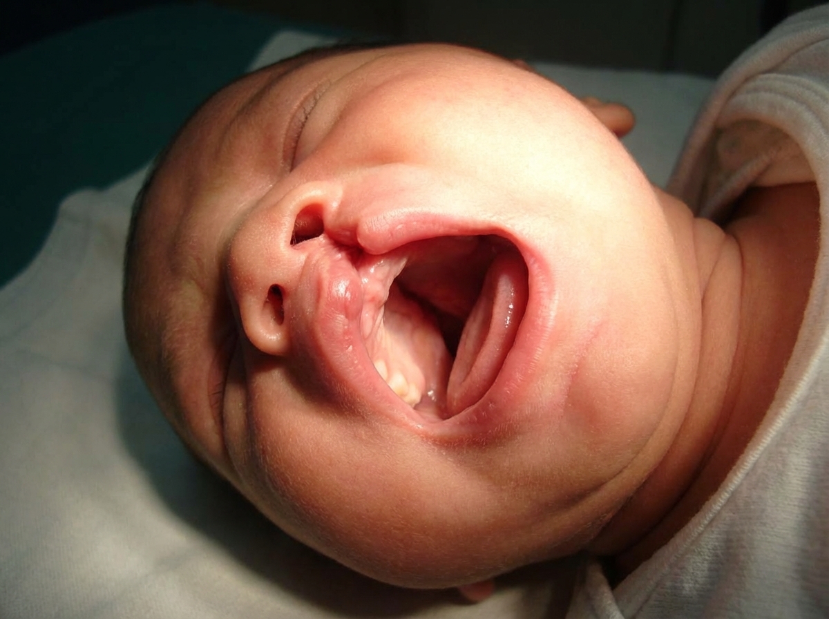
What is the diagnosis of a child presenting with a pulsatile swelling on the medial side of the nose?
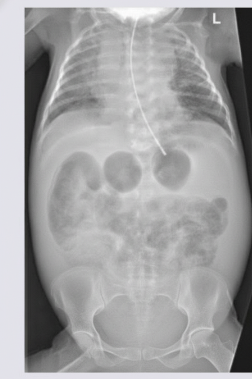
A newborn infant presents with a history of multiple episodes of bilious projectile vomiting. An X-ray of the abdomen was performed. What is the most likely diagnosis?

What is the most likely diagnosis?

Which of the following is NOT true about congenital hypertrophic pyloric stenosis (CHPS)?
What is the most common type of diaphragmatic hernia in children?
What is the commonest cause of intestinal obstruction in a newborn?
What is the most common type of dental caries seen in a primary first molar?
A child underwent splenectomy for splenic injury and blood loss following trauma. What is the recommended further management, considering all EXCEPT which of the following?
True regarding cystic hygroma is:
The covering over an omphalocele is?
Explanation: ### Explanation The correct diagnosis is **Meningocele**. **Why Meningocele is the Correct Answer:** A meningocele is a type of neural tube defect where the meninges herniate through a bony defect in the skull (cranium bifidum). When located at the base of the skull, it can present as a mass on the **medial side of the nose** (nasoethmoidal or nasofrontal). The hallmark clinical feature is that the swelling is **pulsatile** and exhibits an **expansile impulse on coughing or crying** (Furstenberg test). This occurs because the sac is in direct communication with the subarachnoid space, allowing cerebrospinal fluid (CSF) pressure changes to be transmitted to the swelling. **Analysis of Incorrect Options:** * **A. Teratoma:** These are germ cell tumors containing elements from all three germ layers. While they can occur in the head and neck (epignathus), they are typically solid, heterogeneous, and non-pulsatile. * **C. Dermoid Cyst:** This is the most common midline nasal mass. However, dermoid cysts are **non-pulsatile** and do not increase in size with crying because they do not communicate with the intracranial space. They often present with a small pit or sinus tract. * **D. Carcinoma of the ethmoid bone:** This is extremely rare in children. It presents as a destructive, solid, rapidly growing mass, often associated with epistaxis and nasal obstruction, rather than a soft, pulsatile swelling. **High-Yield Clinical Pearls for NEET-PG:** * **Furstenberg Test:** Positive in encephaloceles/meningoceles (compression of internal jugular veins leads to enlargement of the mass). * **Imaging:** MRI is the gold standard to evaluate intracranial extension before any surgical intervention or biopsy. * **Contraindication:** **Never aspirate or biopsy** a midline nasal mass until a meningocele has been ruled out, as this can lead to a CSF leak and meningitis.
Explanation: ***Jejunal atresia*** - **Bilious projectile vomiting** in a newborn with X-ray showing **triple-bubble sign** or **dilated proximal small bowel loops** with **absent distal gas** is characteristic of jejunal atresia. - The obstruction occurs in the **proximal jejunum**, leading to dilation of the stomach, duodenum, and proximal jejunal segment. *Duodenal atresia* - Typically presents with **double-bubble sign** on X-ray, showing dilated stomach and duodenum only. - While it causes **bilious vomiting**, the X-ray pattern differs from the **multiple dilated loops** seen in jejunal atresia. *Ileal atresia* - Characterized by **multiple dilated small bowel loops** throughout the abdomen on X-ray, not the specific triple-bubble pattern. - Occurs more **distally** in the small intestine, allowing more bowel segments to dilate before the obstruction. *Hypertrophic pyloric stenosis* - Presents with **non-bilious projectile vomiting** as the obstruction is **proximal to the ampulla of Vater**. - Diagnosis is typically made by **ultrasound** showing thickened pyloric muscle, not by abdominal X-ray findings.
Explanation: ***Cleft lip and palate*** - The image shows both a **visible cleft in the lip** extending from the nostril downward and a **discontinuity in the hard palate**, indicating involvement of both structures. - This combined defect occurs when **fusion failure** affects both the **primary palate** (lip and anterior alveolus) and **secondary palate** (hard and soft palate) during embryonic development. *Cleft lip* - Would only show a **gap in the lip** without involvement of the **hard or soft palate**. - The **palate would appear intact** on examination, which is not the case here. *Cleft palate* - Would present with **intact lip** but show defects only in the **hard and/or soft palate**. - There would be **no visible external facial deformity**, unlike what is demonstrated in this image. *Bifid uvula* - Represents a **minor form of cleft palate** with only the **uvula being split**. - The **hard palate remains intact** and there is **no lip involvement**, making it a much less severe presentation.
Explanation: ### Explanation: Congenital Hypertrophic Pyloric Stenosis (CHPS) **Why Option D is the Correct Answer:** Ultrasound (USG) is actually the **gold standard** and investigation of choice for diagnosing CHPS. It is highly sensitive (95-100%) and specific. Diagnostic criteria on USG include a **pyloric muscle thickness >3 mm** and a **pyloric canal length >14 mm**. Therefore, stating that USG is "not useful" is factually incorrect. **Analysis of Other Options:** * **Option A (Males commonly affected):** This is true. CHPS shows a strong male predilection, with a male-to-female ratio of **4:1**. It is particularly common in first-born male infants. * **Option B (Presents between 3-8 weeks):** This is true. While symptoms can start as early as 2 weeks, the classic presentation of non-bilious, projectile vomiting occurs between **3 to 6 weeks** of life. * **Option C (X-ray findings):** This is true. A plain abdominal X-ray typically shows a **distended stomach gas shadow** (gastric dilation) with a "caterpillar sign" (hyperperistaltic waves) and a relative **paucity of gas** in the distal small intestines due to the gastric outlet obstruction. **High-Yield Clinical Pearls for NEET-PG:** * **Metabolic Abnormality:** The classic triad is **Hypochloremic, Hypokalemic, Metabolic Alkalosis** with paradoxical aciduria. * **Physical Exam:** A palpable, firm, mobile, ovoid mass in the epigastrium known as the **"Olive-shaped mass"** is pathognomonic. * **Barium Swallow Signs:** If USG is inconclusive, barium studies may show the **"String sign"** (narrowed pyloric canal) or **"Beak sign."** * **Management:** The definitive surgical treatment is **Ramstedt’s Pyloromyotomy**. However, the immediate priority is always the correction of dehydration and electrolyte imbalances.
Explanation: **Explanation:** **Bochdalek Hernia (Correct Answer):** Congenital Diaphragmatic Hernia (CDH) occurs due to the failure of the pleuroperitoneal membranes to fuse with the septum transversum. The **Bochdalek hernia** is the most common type, accounting for approximately **85-90%** of cases. It is located **posterolaterally**, most frequently on the **left side** (80-85%) because the left pleuroperitoneal canal closes later than the right, and the liver provides a protective barrier on the right side. **Incorrect Options:** * **Morgagni Hernia:** This is an anterior retrosternal defect (parasternal). It is much rarer (approx. 2-5%), often asymptomatic in infancy, and usually presents later in life. * **Eventration of the Diaphragm:** This is not a true hernia but a condition where the diaphragmatic muscle is replaced by thin fibroelastic tissue. The diaphragm is intact but elevated, leading to paradoxical movement. * **Hiatus Hernia:** This involves the protrusion of the stomach through the esophageal hiatus. While common in adults, it is a separate clinical entity from the congenital pleuroperitoneal defects seen in neonates. **High-Yield Clinical Pearls for NEET-PG:** * **Triad of CDH:** Cyanosis, Dyspnea, and Dextrocardia (if left-sided). * **Physical Exam:** Scaphoid abdomen and absent breath sounds on the affected side. * **Management:** The primary cause of mortality is **Pulmonary Hypoplasia** and Pulmonary Hypertension, not the defect itself. * **Pro-tip:** Bag-and-mask ventilation is **contraindicated** as it distends the herniated bowel, further compressing the lungs. Immediate endotracheal intubation is the gold standard.
Explanation: **Explanation:** **Duodenal atresia** is the most common cause of congenital intestinal obstruction in newborns. It results from a failure of recanalization of the duodenum during the 8th to 10th week of gestation. Clinically, it presents shortly after birth with **bilious vomiting** (as the obstruction is usually distal to the ampulla of Vater) and epigastric distension. The classic radiographic finding is the **"Double Bubble Sign,"** representing air in the stomach and the proximal duodenum. **Analysis of Options:** * **Annular Pancreas (A):** While it is a common cause of duodenal obstruction, it is less frequent than atresia. It occurs due to the failure of the ventral pancreatic bud to rotate properly, encircling the second part of the duodenum. * **Jejunal Atresia (C):** This is caused by an in-utero **vascular accident** (ischemic necrosis) rather than a recanalization failure. While common, its incidence is lower than that of duodenal atresia. * **Oesophageal Atresia (D):** This presents with drooling, choking, and cyanosis during feeding, but it is an esophageal anomaly, not an *intestinal* obstruction. **High-Yield Clinical Pearls for NEET-PG:** * **Association:** Approximately 30% of infants with duodenal atresia have **Down Syndrome (Trisomy 21)**. * **Antenatal finding:** Maternal **polyhydramnios** is frequently noted due to the fetus's inability to swallow and absorb amniotic fluid. * **Management:** The surgical treatment of choice is **Duodenoduodenostomy** (Diamond-shaped anastomosis). * **Vomiting Rule:** Obstruction proximal to the ampulla of Vater (rare) results in non-bilious vomiting; distal obstruction (common) results in bilious vomiting.
Explanation: ### Explanation In primary molars, the anatomical configuration and the broad, flat nature of the contact areas make them highly susceptible to specific patterns of decay. **Why Option B is Correct:** The most common site for dental caries in primary molars is the **proximal surface**, specifically **below the contact point** (gingival to the contact area). Unlike permanent teeth, which have "point" contacts, primary molars have broad, "surface-to-surface" contacts. This anatomical feature facilitates the entrapment of food debris and plaque, which is difficult to clear with routine brushing. The area just below this contact point is a stagnation zone where acid-producing bacteria (like *Streptococcus mutans*) proliferate, leading to demineralization. **Analysis of Incorrect Options:** * **Option A (Occlusal pits and fissures):** While these are common sites for caries in permanent molars due to deep morphology, in primary teeth, proximal surfaces are statistically more frequently involved, especially as the child ages and the dental arch closes. * **Option C (Proximal surface above the contact point):** The area above the contact point is more accessible to the mechanical cleansing action of chewing and saliva flow, making it less prone to decay than the protected area below the contact. * **Option D (Buccal surface below the height of contour):** This area is generally easier to clean and is more commonly associated with "Smooth Surface Caries" or "Early Childhood Caries" (ECC) related to prolonged bottle feeding, rather than the standard progression of molar decay. **High-Yield Clinical Pearls for NEET-PG:** * **Sequence of Susceptibility:** In primary dentition, the order of caries susceptibility is: Second Molars > First Molars > Maxillary Incisors. * **Early Childhood Caries (ECC):** Characteristically involves the maxillary incisors first; the mandibular incisors are usually spared due to the protective effect of the tongue and submandibular salivary secretions. * **Radiology:** Bitewing radiographs are the gold standard for detecting these proximal lesions, as they are often clinically "hidden" until they become large.
Explanation: **Explanation:** The primary concern following a splenectomy in children is the risk of **Overwhelming Post-Splenectomy Infection (OPSI)** and changes in hematological parameters. **Why Option B is the Correct Answer (The "Except" Option):** Following splenectomy, patients often develop **reactive thrombocytosis** (elevated platelet count) because the spleen is no longer present to sequester and remove aging platelets. While platelet counts can rise significantly, this is usually a transient, physiological response and rarely leads to spontaneous thromboembolic events in children. Therefore, **routine administration of aspirin** (an antiplatelet agent) is **not recommended** unless the platelet count exceeds extremely high thresholds (typically >1.5 million/µL) or the patient has additional prothrombotic risk factors. **Analysis of Other Options:** * **Option A (Continuous Penicillin):** This is standard practice. Children who have undergone splenectomy require daily prophylactic antibiotics (usually Penicillin V) to prevent OPSI, particularly against encapsulated organisms like *Streptococcus pneumoniae*. This is often continued until at least age 5 or for at least 2–5 years post-surgery. * **Option C & D (Education and Evaluation):** Parental education is critical. They must be warned that any fever in a splenectomized child is a medical emergency requiring immediate evaluation and broad-spectrum antibiotics to prevent rapid sepsis. **NEET-PG High-Yield Pearls:** * **OPSI Risk:** Highest in the first 2 years post-splenectomy; most common organism is *S. pneumoniae*. * **Vaccination Protocol:** Ideally, vaccines (Pneumococcal, Meningococcal, *H. influenzae* type b) should be given **14 days before** elective surgery or **14 days after** emergency surgery. * **Peripheral Smear Findings:** Look for **Howell-Jolly bodies**, Pappenheimer bodies, and Heinz bodies post-splenectomy. * **Splenic Salvage:** In pediatric trauma, the current "gold standard" is non-operative management to preserve splenic immune function.
Explanation: **Explanation:** **Cystic Hygroma** (also known as macrocystic lymphatic malformation) is a congenital malformation of the lymphatic system, resulting from the failure of lymphatics to connect with the venous system. 1. **Why Option B is correct:** Cystic hygromas are thin-walled, multiloculated cysts filled with clear, straw-colored lymph fluid. Because they contain clear fluid and lack a solid component or thick capsule, they are **brilliantly translucent** when a light source is applied (transillumination test). This is a classic clinical sign used to differentiate them from other neck masses like hemangiomas or teratomas. 2. **Why other options are incorrect:** * **Option A:** While most commonly found in the posterior triangle of the neck, they can be bilateral or extend across the midline. * **Option C:** Unlike strawberry hemangiomas, cystic hygromas **do not spontaneously regress**. They typically grow in proportion to the child and require intervention (sclerotherapy or surgical excision) if they cause functional impairment or cosmetic deformity. * **Option D:** Although 50–60% are present at birth, about 90% manifest by age two. They can appear later in life following trauma or infection. **High-Yield Clinical Pearls for NEET-PG:** * **Most common site:** Left side of the neck (Posterior triangle). * **Association:** Frequently associated with chromosomal anomalies, most notably **Turner Syndrome (45, XO)** and Down Syndrome. * **Complications:** Sudden increase in size usually indicates **intracystic hemorrhage** or infection. * **Management:** **Sclerotherapy** (e.g., OK-432, Bleomycin) is often the first-line treatment for macrocystic lesions; surgical excision is reserved for cases where sclerotherapy fails.
Explanation: **Explanation:** An **omphalocele** is a midline ventral abdominal wall defect at the base of the umbilical cord. The hallmark of this condition is that the herniated viscera (usually bowel and sometimes liver) are contained within a protective sac. **Why the correct answer is right:** The omphalocele sac is a trilaminar structure. From the inside out, it consists of **peritoneum**, a middle layer of **Wharton’s jelly**, and an outer layer of **amniotic membrane**. The umbilical cord inserts directly into the apex of this sac, rather than the abdominal wall. **Why the incorrect options are wrong:** * **Skin:** In a true omphalocele, the skin stops at the edge of the defect and does not cover the herniated contents. If the defect were covered by skin, it would be classified as an umbilical hernia. * **Chorionic membrane:** The chorion is the outer fetal membrane that contributes to the placenta. It does not play a role in covering abdominal wall defects; the amnion is the membrane in direct contact with the fetus and the umbilical cord. **High-Yield Clinical Pearls for NEET-PG:** * **Omphalocele vs. Gastroschisis:** Unlike omphalocele, gastroschisis occurs to the *right* of the umbilical cord and has **no covering sac** (the bowel is exposed to amniotic fluid, leading to chemical peritonitis). * **Associated Anomalies:** Omphalocele is highly associated with chromosomal abnormalities (Trisomy 13, 18, 21) and syndromes like **Beckwith-Wiedemann Syndrome** (macroglossia, gigantism, hypoglycemia). * **Management:** Small defects can be closed primarily. Large ("Giant") omphaloceles may require staged closure using a "silo" or conservative management with escharotic agents (e.g., silver sulfadiazine) to allow the sac to epithelialize.
Surgical Conditions of the Newborn
Practice Questions
Congenital Diaphragmatic Hernia
Practice Questions
Esophageal Atresia and Tracheoesophageal Fistula
Practice Questions
Intestinal Atresia and Stenosis
Practice Questions
Malrotation and Volvulus
Practice Questions
Hirschsprung's Disease
Practice Questions
Anorectal Malformations
Practice Questions
Biliary Atresia
Practice Questions
Abdominal Wall Defects
Practice Questions
Inguinal Hernia and Hydrocele
Practice Questions
Intussusception
Practice Questions
Appendicitis in Children
Practice Questions
Get full access to all questions, explanations, and performance tracking.
Start For Free