In infants and toddlers, craniotabes is a sign related to the deficiency of
Among the following, the best indicator for acute malnutrition in the under-fives is
Which of the following criteria are used for detecting a child with severe acute malnutrition according to WHO guidelines? 1. Bilateral pitting oedema 2. Weight for height Z-score less than minus three SD 3. Mid upper arm circumference of less than 11.5 cm (115 mm) Select the correct answer using the code given below:
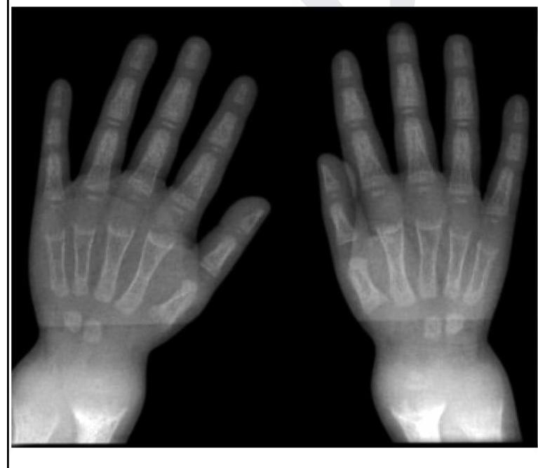
A child presents with poor growth and swelling at joints. A radiograph of his wrist is given below. Lab investigations reveal serum ALP levels of >1500. What is the possible diagnosis?

Dose of vitamin A for an 18 month old baby, with keratomalacia, weighing 10 kg is?
Which is the most specific clinical feature for diagnosis of Kwashiorkor?
For severe acute malnutrition in children (6-59 months), MAC will be less than
In marasmus, which of the following is characteristically seen?
A breast fed child presents with hypernatremia (Serum sodium > 170m Eq/L). His urine sodium is 70 mEq/L. Which of the following is the most likely cause –
Craniotabes is seen in
Explanation: ***Vitamin D (Correct Answer)*** - **Craniotabes** refers to the softening of the cranial bones, characterized by a "ping-pong ball" sensation on palpation. - It is an **early clinical sign of rickets** in infants, which results from **vitamin D deficiency**. - **Vitamin D** is essential for the absorption and metabolism of **calcium and phosphate**, which are critical for proper bone mineralization. - Without adequate vitamin D, bones remain inadequately mineralized, leading to softening. *Vitamin K (Incorrect)* - **Vitamin K** plays a role in **blood coagulation** (clotting factors II, VII, IX, X) and bone metabolism through carboxylation of osteocalcin. - Deficiency manifests primarily as **bleeding disorders** (hemorrhagic disease of newborn), not skeletal abnormalities. - Does not cause craniotabes or bone softening. *Vitamin A (Incorrect)* - **Vitamin A** is essential for **vision** (rhodopsin formation), **immune function**, and **epithelial cell differentiation**. - Deficiency causes **night blindness** (earliest sign), **xerophthalmia**, **Bitot's spots**, and increased susceptibility to infections. - Not associated with skeletal manifestations like craniotabes. *Vitamin C (Incorrect)* - **Vitamin C** (ascorbic acid) is required for **collagen synthesis** (hydroxylation of proline and lysine). - Deficiency causes **scurvy**, characterized by **bleeding gums**, **petechiae**, **subperiosteal hemorrhages**, and impaired wound healing. - While scurvy affects bone matrix and periosteum, it does not cause the characteristic softening of cranial bones seen in craniotabes.
Explanation: ***Weight for height*** - **Weight-for-height** is the best indicator for **acute malnutrition** (wasting) in under-fives as it reflects recent nutritional deficits. - It compares a child's weight to the expected weight for a child of the same height, identifying if they are too thin for their height. *Height for age* - **Height-for-age** is an indicator of **chronic malnutrition (stunting)**, reflecting long-term nutritional deprivation. - It does not accurately capture acute, recent weight loss or wasting. *Head/chest circumference ratio* - The **head/chest circumference ratio** can be used as a screening tool in some contexts, but it is less precise and sensitive for assessing acute malnutrition than weight-for-height. - Its utility decreases beyond the first year of life as the chest circumference typically begins to exceed head circumference. *Mid arm circumference* - **Mid-upper arm circumference (MUAC)** is a useful **screening tool** for severe acute malnutrition, particularly in community settings. - However, **weight-for-height** is generally considered a more comprehensive and accurate indicator for diagnosing and assessing the severity of acute malnutrition across all severities.
Explanation: ***1, 2 and 3*** - According to **WHO guidelines**, Severe Acute Malnutrition (SAM) in children aged 6-59 months is diagnosed by the presence of **ANY ONE** of these three criteria: - **Bilateral pitting oedema** (nutritional edema) - indicates kwashiorkor - **Weight-for-height Z-score (WHZ) < -3 SD** - indicates severe wasting - **Mid-Upper Arm Circumference (MUAC) < 11.5 cm (115 mm)** - indicates severe wasting - All three are **independent diagnostic criteria** for SAM, meaning any single criterion is sufficient for diagnosis - These criteria are used in **community screening**, **facility-based assessment**, and **nutritional rehabilitation programs** *1 and 2 only* - This incorrectly excludes **MUAC < 11.5 cm**, which is a valid and widely used WHO criterion for SAM diagnosis - MUAC is particularly useful for **community-based screening** as it's simple and doesn't require complex equipment *2 and 3 only* - This incorrectly excludes **bilateral pitting oedema**, which is a critical criterion for diagnosing **kwashiorkor** (edematous malnutrition) - Oedema can occur even when weight-for-height appears normal due to fluid retention masking tissue wasting *1 and 3 only* - This incorrectly excludes **weight-for-height Z-score < -3 SD**, which is the **gold standard anthropometric measure** for severe wasting - WHZ is essential for facility-based diagnosis and monitoring treatment response
Explanation: ***Rickets*** - The combination of **poor growth**, **joint swelling**, and **elevated alkaline phosphatase (ALP)** in a child strongly indicates rickets, a condition of defective bone mineralization in growing bones. - The radiograph of the wrist would likely show typical findings like **widened growth plates**, **fraying** and **cupping of metaphyses**, and **decreased bone density**, which are characteristic of rickets. *Osteoporosis* - This condition is characterized by **reduced bone mass** and **fragile bones**, typically seen in older adults or due to secondary causes, and is not primarily linked to joint swelling in children. - While ALP levels can be normal or slightly elevated in osteoporosis, a level of >1500 is highly suggestive of active bone formation or breakdown, not typically seen in osteoporosis. *Osteomalacia* - This is defective bone mineralization in adults after growth plates have fused, leading to **bone softening** and **pain**, typically not presenting with joint swelling as a primary symptom. - While it also involves high ALP and bone demineralization, the clinical context of a *child* with growth issues points more specifically to rickets. *Osteogenesis imperfecta* - This is a group of **genetic disorders** characterized by **brittle bones** that fracture easily, often accompanied by **blue sclerae** and **hearing loss**, which are not mentioned in the presentation. - While bone fragility is present, it does not typically cause the described joint swelling or the significantly elevated ALP levels seen in this case.
Explanation: **2,00,000 IU** - For children 12 months of age and older with **keratomalacia** due to vitamin A deficiency, the recommended dose is **200,000 IU** orally, given immediately. - This dose should be repeated the next day and again after four weeks to replenish stores and prevent recurrence. *1,00,000 IU* - This dose is typically recommended for infants **aged 6 to 11 months** with **clinical vitamin A deficiency**, including keratomalacia. - It is insufficient for an 18-month-old child with active keratomalacia. *50,000 IU* - This dose is usually given to infants **under 6 months** of age with clinical signs of **vitamin A deficiency**. - It is too low for an 18-month-old baby with keratomalacia. *5,00,000 IU* - This dose is excessively high and potentially toxic for an 18-month-old child. - Vitamin A toxicity can lead to adverse effects, including **increased intracranial pressure** and liver damage.
Explanation: ***Easy pluckable hair*** - **Easy pluckable hair** is a highly specific clinical sign of Kwashiorkor, indicating severe **protein deficiency** affecting hair follicle integrity. - This symptom, often accompanied by changes in hair color and texture (e.g., **flag sign**), highlights the chronic lack of essential amino acids. *Edema* - **Edema** is a prominent feature of Kwashiorkor, resulting from low **oncotic pressure** due to **hypoalbuminemia**. - While characteristic, edema can also be seen in other conditions like **cardiac**, **renal**, or **hepatic failure**, making it less specific than hair changes. *Fatty liver* - **Fatty liver** (hepatic steatosis) is commonly observed in Kwashiorkor due to impaired synthesis and export of **VLDL** from the liver. - Although characteristic, fatty liver can also occur in **obesity**, **alcoholism**, and **diabetes**, reducing its specificity as a standalone diagnostic feature. *Low serum albumin* - **Low serum albumin** is a hallmark biochemical finding in Kwashiorkor, reflecting severe **protein malnutrition** and reduced hepatic synthesis. - While critical for diagnosis, hypoalbuminemia can also be caused by severe **liver disease**, **nephrotic syndrome**, or **malabsorption**, limiting its specificity for Kwashiorkor alone.
Explanation: ***11.5 cm*** - A **Mid-Upper Arm circumference (MUAC) below 11.5 cm** is a key diagnostic criterion for **severe acute malnutrition (SAM)** in children aged 6-59 months. - This measurement is a simple and effective screening tool in resource-limited settings to identify children at high risk of mortality due to malnutrition [1]. *13.5 cm* - A MUAC of 13.5 cm or greater is generally considered **nutritionally healthy** for children in this age group, indicating adequate muscle and fat reserves. - This measurement would typically rule out severe acute malnutrition and often even moderate malnutrition. *12.5 cm* - A MUAC between 11.5 cm and 12.5 cm is typically indicative of **moderate acute malnutrition (MAM)**, not severe acute malnutrition. - While concerning, it suggests a less critical nutritional status compared to a MUAC below 11.5 cm. *14.5 cm* - A MUAC of 14.5 cm or greater is well within the healthy range for children aged 6-59 months, indicating **good nutritional status**. - This measurement would suggest no signs of acute malnutrition.
Explanation: ***Severe muscle wasting*** - **Severe muscle wasting** and **loss of subcutaneous fat** are hallmark features of marasmus, giving the child a characteristic **"skin and bones"** appearance. - Children present with **visible ribs**, **sunken cheeks**, and **prominent bony landmarks** due to depletion of both fat and muscle stores from chronic energy deficiency. *Hepatomegaly* - **Hepatomegaly** is a characteristic feature of **kwashiorkor**, not marasmus, caused by **fatty infiltration** of the liver due to impaired lipoprotein synthesis. - In marasmus, the liver is typically **normal or reduced in size** as fat stores are mobilized and utilized for energy needs. *Edema* - **Pitting edema** is a hallmark feature of **kwashiorkor**, resulting from severe protein deficiency causing **hypoalbuminemia** and reduced plasma oncotic pressure. - Marasmus is characterized by **absence of edema** because the protein deficiency is proportionally less severe compared to the overall caloric deficiency. *Voracious appetite* - Children with marasmus typically have **poor appetite** and **feeding difficulties**, not increased hunger, due to severe weakness and apathy. - The **energy depletion** and **muscle wasting** significantly reduce the child's interest in food and ability to consume adequate amounts.
Explanation: ***Excessive intake of sodium*** - A critically elevated **serum sodium (>170 mEq/L)** coupled with a high **urine sodium (70 mEq/L)** in a breastfed infant indicates that the kidneys are actively trying to excrete excess sodium. This pattern is consistent with an exogenous sodium overload. - This scenario suggests the ingestion of a **hypertonic solution** or food, likely by mistake, leading to significant sodium toxicity requiring rapid renal excretion. *Acute tubular necrosis* - In ATN, there's impaired renal concentration and reabsorption, but acute kidney injury often leads to **normonatremia or hyponatremia**, not severe hypernatremia. - While urine sodium can be high in ATN due to tubular damage, the primary cause of such extreme hypernatremia would typically be external sodium load. *Severe dehydration* - Severe dehydration usually causes **pre-renal acute kidney injury**, characterized by **high serum sodium** due to water loss, but the kidneys would **conserve sodium**, resulting in a very **low urine sodium** (<20 mEq/L). - The high urine sodium of 70 mEq/L in this case **argues against dehydration** as the primary cause of hypernatremia. *Diabetes insipidus* - Diabetes insipidus (DI) causes **hypernatremia due to free water loss** from the kidneys, resulting in a **dilute urine** with a **low urine osmolality** and typically **low urine sodium**. - The elevated urine sodium of 70 mEq/L is inconsistent with the renal handling of sodium seen in diabetes insipidus.
Explanation: ***Rickets*** - **Craniotabes**, characterized by softening of the skull bones, is the **classic and most common** sign of **rickets** due to defective **bone mineralization**. - It is frequently seen in infants with **vitamin D deficiency**, leading to uncalcified skull bones that can be indented on gentle pressure. - This is the **expected answer** for craniotabes in medical examinations. *Hydrocephalus* - While hydrocephalus can cause an **enlarged head** and increased intracranial pressure, it does not directly cause the softening or **craniotabes** of the skull bones. - The skull bones might separate at the sutures due to pressure, but they don't become soft and compressible in the typical pattern of craniotabes. *Osteogenesis imperfecta* - This is a genetic disorder of **type I collagen** causing **brittle bones** that fracture easily. - While defective bone formation can rarely present with skull softening, craniotabes is **not a characteristic feature** of this condition. - Classic features include **blue sclerae**, **dentinogenesis imperfecta**, and **multiple fractures**. *All of the options* - This is incorrect because **craniotabes** is **characteristically and classically** associated with **rickets**, which is the primary pathological cause. - While other conditions may affect bone structure, rickets remains the definitive answer for craniotabes in clinical practice.
Breastfeeding
Practice Questions
Infant Formula Feeding
Practice Questions
Complementary Feeding
Practice Questions
Nutritional Requirements by Age
Practice Questions
Malnutrition and Failure to Thrive
Practice Questions
Obesity in Children
Practice Questions
Vitamin Deficiencies
Practice Questions
Mineral Deficiencies
Practice Questions
Food Allergies and Intolerances
Practice Questions
Nutritional Management of Chronic Diseases
Practice Questions
Eating Disorders
Practice Questions
Parenteral and Enteral Nutrition
Practice Questions
Get full access to all questions, explanations, and performance tracking.
Start For Free