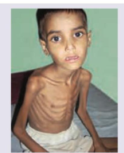
All statements about the disease shown are wrong except:

Select the false statement regarding the disease depicted in the picture?

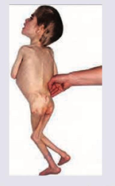
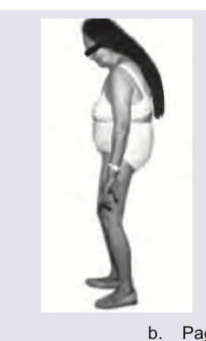
All are true about the child shown in the image except:

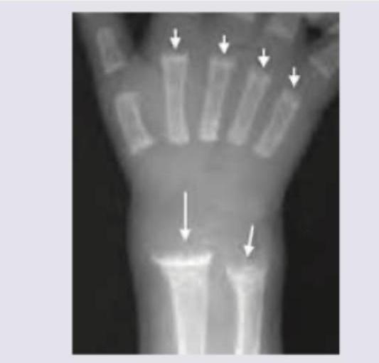
A 2-year-old child presents with delayed motor milestones and bowing of legs. X-ray of the wrist is shown. What is the most likely diagnosis?

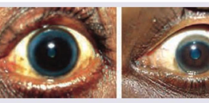
Which condition is characterized by the sign shown in the image?

The image shows:

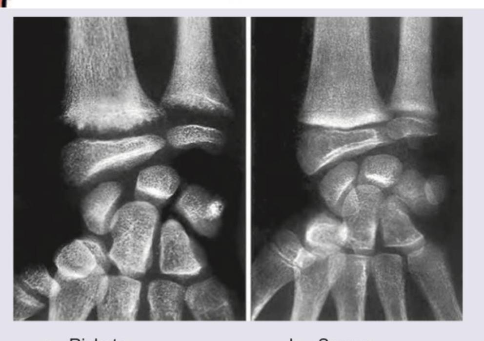
Which of the following is correct about the image shown? (Recent NEET Pattern 2016-17)

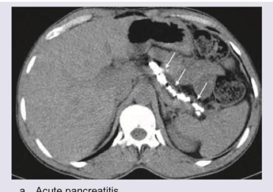
Which of the following diseases will lead to the following appearance?

The bony deformity of 'pigeon chest' in children occurs due to deficiency of:
In the case of a 7 -year-old school-going child, which would be the most appropriate indicator to measure the current nutritional status?
Explanation: **This is an EXCEPT question** - three statements are WRONG, one is CORRECT. ***Loss of normal zone of provisional calcification adjacent to metaphysis*** ✓ **CORRECT STATEMENT** - This is a **true statement** and the correct answer to this EXCEPT question. - The image shows findings consistent with **rickets**, characterized by **widened, irregular growth plates** at the wrists. - The **zone of provisional calcification** is where cartilage is normally mineralized before ossification; its loss or irregularity is a **hallmark radiological finding of rickets**. - This occurs due to inadequate calcium and phosphorus for proper mineralization. *Nutritional rickets usually presents in school age, usually as widened wrists* - **WRONG STATEMENT** - This statement is **FALSE** (making it incorrect for this EXCEPT question). - Nutritional rickets typically manifests in **infancy or early childhood (3 months to 3 years)**, NOT school age. - While widened wrists are indeed a clinical feature, the age of presentation stated here is incorrect. - School-age presentation is more typical of **X-linked hypophosphatemic rickets**, not nutritional rickets. *Presentation in early infancy along with seizures or tetany* - **WRONG STATEMENT** - This statement is **FALSE** (making it incorrect for this EXCEPT question). - While seizures or tetany CAN occur in rickets due to **severe hypocalcemia**, they represent **complications**, not typical initial presentations. - Typical early presentations include **craniotabes** (skull softening), delayed fontanelle closure, frontal bossing, and rachitic rosary. - Seizures/tetany occur only in severe cases with marked hypocalcemia. *Excessive exposure to sunlight does not increase vitamin D production* - **WRONG STATEMENT** - This statement is **FALSE** (making it incorrect for this EXCEPT question). - Sunlight exposure **DOES increase vitamin D production** - this is well-established physiology. - **UVB radiation** converts 7-dehydrocholesterol in skin to previtamin D3, which isomerizes to vitamin D3. - Sunlight is the **primary natural source** of vitamin D synthesis in humans.
Explanation: ***Loss of fat from sucking pads of the cheeks is the earliest sign*** - The **Buccal fat pad** (sucking pads of the cheeks) tends to be one of the **last fat deposits** to be mobilized in severe starvation, making their loss one of the later signs of marasmus, not the earliest. - Earlier signs of fat loss are typically observed in the **axillary** and **gluteal regions**, and then the **limbs** and **trunk**. *There is muscle atrophy and resultant hypotonia* - In **marasmus**, there is severe **wasting of muscle tissue** and subcutaneous fat due to a chronic deficiency of both energy and protein. - This muscle wasting leads to a significant decrease in muscle mass and tone, resulting in generalized **hypotonia**. *Besan panjin is an energy dense food used in treatment* - **Besan panjiri** powder, made from roasted gram flour (besan), sugar, and ghee, is a traditional Indian **energy-dense food** often used in nutritional rehabilitation for severely malnourished children. - It provides a good source of **calories** and **protein** essential for recovery in conditions like marasmus. *This is non-edematous severe childhood undernutrition* - The image shows a child with extreme wasting and absence of edema, which is characteristic of **marasmus**. - **Marasmus** is a form of **severe acute malnutrition** (SAM) that is typically non-edematous, distinguishing it from kwashiorkor.
Explanation: ***Delayed skin pinch*** - Delayed skin pinch, also known as **poor skin turgor**, is a sign of **dehydration**, which is more characteristic of **kwashiorkor** or severe acute malnutrition with significant fluid loss, rather than marasmus alone. - In marasmus, although there is severe wasting, **skin turgor is often preserved** due to the absence of significant edema. - This is the **EXCEPTION** - it is NOT a typical feature of pure marasmus. *Baggy pants appearance* - The **extreme loss of subcutaneous fat and muscle mass** in marasmus causes the skin around the buttocks and thighs to hang loosely, creating a "baggy pants" or "old man" appearance. - This is a **hallmark feature** indicating severe wasting in marasmus. *Apathy* - Children with severe marasmus often exhibit **apathy, irritability, and decreased activity**, reflecting their profound energy deficit and associated psychological distress. - This is a common neurological and behavioral manifestation of **severe malnutrition**. *Reduced MAC* - **Mid-upper arm circumference (MAC)** is severely reduced in marasmus due to the significant loss of both fat and muscle tissue in the upper arm. - A **MAC less than 11.5 cm** is a key diagnostic criterion for **severe acute malnutrition** in children aged 6-59 months.
Explanation: ***Rickets*** - The image displays characteristic radiographic findings of **rickets**, including **fraying, cupping**, and **widening of the metaphyses**, particularly evident at the distal radius and ulna. - These changes result from impaired mineralization of growing bone and cartilage at the **growth plates** due to vitamin D deficiency in children. *Scurvy* - Scurvy, caused by vitamin C deficiency, affects **collagen synthesis**, leading to subperiosteal hemorrhages and thinning of the cortex. - Radiographic features include a **dense metaphyseal line (Wimberger's sign)** and **epiphyseal separation**, which are not the primary features seen here. *Osteomalacia* - **Osteomalacia** is the adult form of rickets, characterized by defective mineralization of bone matrix. - While it features **pseudofractures (Looser zones)** and generalized osteopenia, it does not present with growth plate abnormalities like fraying and cupping, as growth plates are typically closed in adults. *Osteoporosis* - **Osteoporosis** involves a reduction in bone mass and density, leading to increased bone fragility and fracture risk. - Radiographically, it manifests as generalized **osteopenia** and trabecular thinning, without the specific metaphyseal irregularities seen in rickets.
Explanation: ***Kwashiorkor*** - The image shows sparse, dry, and discolored hair, sometimes described as having a **"flag sign"** due to alternating bands of light and dark hair, which is characteristic of **protein-energy malnutrition (PEM)**, specifically kwashiorkor. - Kwashiorkor results from a severe protein deficiency with relatively adequate calorie intake, leading to hair changes, **edema**, and a distended abdomen. *Marasmus* - Marasmus is characterized by severe muscle wasting and loss of subcutaneous fat due to a deficiency in both protein and overall calorie intake, but **hair changes are less prominent** than in kwashiorkor. - Individuals with marasmus appear incredibly emaciated and "skeletal" but typically do not exhibit the same degree of hair discoloration or edema. *Pellagra* - Pellagra is caused by a deficiency of **niacin (vitamin B3)** and is characterized by the "3 Ds": **dermatitis**, **diarrhea**, and **dementia**. - While it can affect hair quality, it does not typically present with the specific hair changes, such as the "flag sign," seen in the image. *None of above* - The distinct hair abnormalities depicted in the image are highly suggestive of a specific nutritional deficiency, making "None of the above" incorrect.
Explanation: ***Acute pancreatitis*** - The image shows an **enlarged, edematous pancreas** with surrounding **stranding of the peripancreatic fat**, indicated by the white arrows and suggesting inflammation. - There is also evidence of **fluid collection** adjacent to the pancreas, a common finding in acute pancreatitis. *Chronic pancreatitis* - Chronic pancreatitis typically presents with pancreatic **atrophy**, **calcifications** within the parenchyma and ducts, and **ductal dilation**. - While calcifications are present, the primary findings of a swollen pancreas and peripancreatic edema are more indicative of an acute process. *Pancreatic divisum* - Pancreatic divisum is a **congenital anomaly** where the dorsal and ventral pancreatic ducts fail to fuse, and it is a predisposing factor for pancreatitis, but the image shows findings of an inflammatory process rather than an anatomical variant. - The image exhibits signs of inflammation and fluid collection, not the distinct ductal anatomy of pancreatic divisum itself. *Artefact* - Artefacts on a CT scan can include **beam hardening**, **motion artefacts**, or **streak artefacts**, which typically appear as streaks, distorted images, or areas of abnormal signal. - The observed features—pancreatic enlargement, peripancreatic fat stranding, and fluid collection—are clear anatomical and pathological changes, not image anomalies.
Explanation: ***A = Kayser Fleischer ring, B = Arcus senilis*** - Image A displays a **Kayser-Fleischer ring**, a greenish-brown ring in the periphery of the cornea, characteristic of **Wilson's disease** due to copper deposition. - Image B shows **arcus senilis**, a milky-white or grayish ring around the cornea, which is a **common age-related change** due to lipid deposits, and can appear at any age. *A = Arcus senilis, B = Kayser-Fleischer ring* - This option incorrectly identifies the lesions; the ring in image A is associated with copper deposition, not lipid deposition. - The ring in image B is clearly a common lipid ring often seen in older individuals, contrasting with the distinct appearance of a Kayser-Fleischer ring. *A = Arcus senilis, B = Hudson-Stahli line* - Image A does not show arcus senilis; its color and location are consistent with copper deposition. - Image B does not depict a Hudson-Stahli line, which is a thin, brownish-linear deposit of iron in the corneal epithelium, typically horizontal and not circumferential. *A = Hudson-Stahli line, B = Arcus senilis* - Image A's appearance, as a circumferential greenish-brown deposit, is not consistent with a linear Hudson-Stahli line. - While image B correctly identifies arcus senilis, the identification of A as a Hudson-Stahli line is incorrect.
Explanation: ***Paget's disease*** - The image depicts a bowing of the legs and a generalized stooped posture, which are characteristic findings in advanced **Paget's disease of bone**. - **Paget's disease** is a chronic bone disorder characterized by abnormal bone remodeling, leading to enlarged and misshapen bones that can cause **kyphosis** and **bowed limbs**. *Polio* - **Polio** (poliomyelitis) primarily affects the nervous system, leading to **muscle weakness** and **paralysis**, which can result in limb deformities, but not typically the widespread bone enlargement and bowing seen here. - While polio can cause limb asymmetry and deformities due to muscle wasting, it does not involve the characteristic bone remodeling and generalized skeletal changes seen in the image. *Cerebellar lesion* - A **cerebellar lesion** primarily affects coordination, balance, and motor control, leading to ataxia, tremors, and gait disturbances. - It does not directly cause the skeletal deformities or bone bowing illustrated in the image. *Ankylosing Spondylitis* - **Ankylosing spondylitis** is a chronic inflammatory disease primarily affecting the axial skeleton, leading to progressive spinal stiffness and fusion (a "bamboo spine"). - While it can cause a significant **kyphosis** (forward curvature of the spine), it does not typically lead to the wide-set bowed legs or general bone enlargement often seen in Paget's disease.
Explanation: ***Vitamin D*** - A deficiency in **Vitamin D** leads to **rickets** in children, impairing proper bone mineralization. - This softening of bones can cause skeletal deformities, including **pigeon chest** (pectus carinatum), where the sternum protrudes forward. *Vitamin K* - **Vitamin K** is crucial for **blood clotting** by synthesizing coagulation factors. - Its deficiency primarily leads to bleeding disorders, not skeletal deformities like pigeon chest. *Vitamin A* - **Vitamin A** is essential for **vision**, immune function, and cellular growth. - A deficiency can cause **night blindness** and impaired immunity, but it is not directly linked to bone deformities. *Vitamin E* - **Vitamin E** is a powerful **antioxidant** that protects cells from damage. - While essential for neurological function and immunity, its deficiency does not cause skeletal abnormalities such as pigeon chest.
Explanation: ***Mid upper arm circumference*** - **Mid-upper arm circumference (MUAC)** is the most appropriate indicator among the given options for assessing **current nutritional status** in a 7-year-old school-going child. - While traditionally emphasized for children 6-59 months, **MUAC is increasingly recognized as a valid indicator for school-aged children (5-15 years)** for detecting acute malnutrition and wasting. - MUAC is **age-independent, practical, and can be measured easily** in school settings, making it particularly useful for screening current nutritional status in this age group. - **Note:** Ideally, **BMI-for-age** is the gold standard recommended by WHO and IAP for children aged 5-19 years, but it is not among the options provided. *Weight for height* - **Weight-for-height (WFH)** is primarily recommended for **children under 5 years of age** or those with height <120 cm according to WHO guidelines. - For school-aged children (>5 years), **BMI-for-age is the preferred indicator**, not WFH. - WFH becomes less accurate and less practical in older children, making it inappropriate as the primary indicator for a 7-year-old. *Birth weight* - **Birth weight** reflects **intrauterine growth and nutritional status at delivery**, not the current nutritional status of a 7-year-old child. - It is useful for assessing risk factors and early life influences but has no bearing on current nutritional assessment in school-aged children. *Head circumference* - **Head circumference** is primarily used to assess **brain growth** and detect conditions like **microcephaly or macrocephaly**, particularly in infancy and early childhood (up to 2-3 years). - In a 7-year-old, head growth has largely plateaued, and this measurement is **not useful for assessing current general nutritional status**.
Breastfeeding
Practice Questions
Infant Formula Feeding
Practice Questions
Complementary Feeding
Practice Questions
Nutritional Requirements by Age
Practice Questions
Malnutrition and Failure to Thrive
Practice Questions
Obesity in Children
Practice Questions
Vitamin Deficiencies
Practice Questions
Mineral Deficiencies
Practice Questions
Food Allergies and Intolerances
Practice Questions
Nutritional Management of Chronic Diseases
Practice Questions
Eating Disorders
Practice Questions
Parenteral and Enteral Nutrition
Practice Questions
Get full access to all questions, explanations, and performance tracking.
Start For Free