In a child, which of the following conditions does NOT typically involve CSF examination for diagnosis?
What is the most common abdominal tumor in children?
A 4-year-old child presented with a palpable abdominal mass in the right flank region which was painless and slowly increasing in size along with some episodes of fever and hematuria. On examination, hypertension was noted. CT scan of the abdomen was done. The patient was operated and the mass was resected. The gross specimen and the HPE examination are given below. All of the following genes can be mutated in the above disease except?

A malignant tumor of childhood that frequently metastasizes to bone marrow most often is:
Neuroblastoma differs from Wilms tumor by all, EXCEPT:
The following physical appearance is seen in which of the following conditions?

The most common site of neuroblastoma is
A 9-year-old female child presents with a history of headache and visual disturbances. What is the most likely diagnosis?
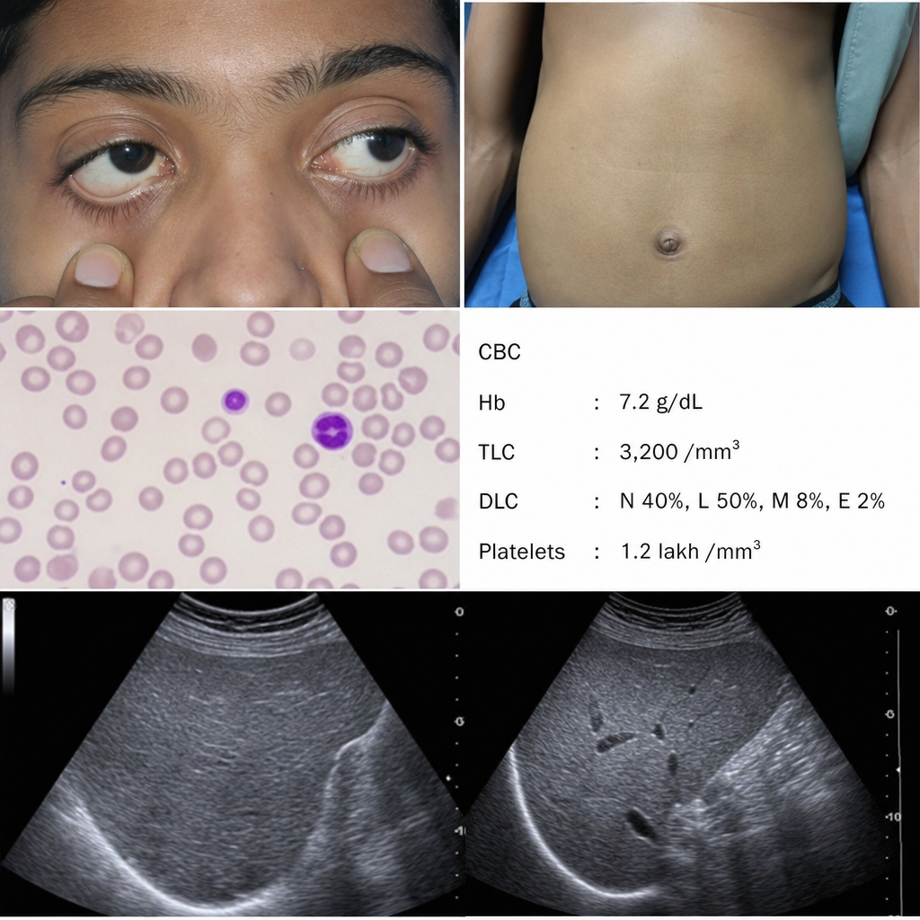
Which of the following is true about leukemia in Down's syndrome?
Which of the following is NOT a good prognostic factor for Acute Lymphoblastic Leukemia (ALL)?
Explanation: **Explanation:** The correct answer is **Hodgkin’s Lymphoma (HL)**. The decision to perform a cerebrospinal fluid (CSF) examination in pediatric oncology is based on the propensity of the malignancy to involve the Central Nervous System (CNS). **1. Why Hodgkin’s Lymphoma is the correct answer:** Hodgkin’s Lymphoma is primarily a disease of the lymph nodes and rarely involves the CNS at the time of diagnosis or during relapse. Because CNS involvement is extremely uncommon, routine lumbar puncture for CSF cytology is not part of the standard staging or diagnostic workup for HL. **2. Why the other options are incorrect:** * **ALL (Acute Lymphoblastic Leukemia):** CNS involvement is common in ALL. CSF examination is mandatory at diagnosis for staging (CNS-1, 2, or 3) and for the administration of intrathecal chemotherapy, which is a cornerstone of treatment. * **AML (Acute Myeloid Leukemia):** While less frequent than in ALL, CNS involvement occurs in approximately 5-10% of pediatric AML cases (especially the monocytic subtypes M4 and M5). Routine CSF analysis is required at diagnosis. * **Non-Hodgkin’s Lymphoma (NHL):** Pediatric NHL (such as Burkitt lymphoma or Lymphoblastic lymphoma) is highly aggressive and has a high predilection for CNS spread. Staging always includes a CSF examination. **Clinical Pearls for NEET-PG:** * **CNS Sanctuary Sites:** The CNS and testes are considered "sanctuary sites" in leukemias because systemic chemotherapy often fails to reach therapeutic concentrations there due to the blood-brain barrier. * **L-Asparaginase:** While used in ALL, it does not cross the BBB; hence, direct intrathecal therapy (Methotrexate, Cytarabine, Hydrocortisone) is vital. * **Staging:** Remember that while HL uses the **Ann Arbor Staging**, pediatric NHL often uses the **St. Jude (Murphy) Staging System**, where any CNS involvement automatically classifies the disease as Stage IV.
Explanation: **Explanation:** **Wilms Tumor (Nephroblastoma)** is the most common primary renal tumor and the **most common abdominal tumor** in children. It typically presents as a large, smooth, firm, and asymptomatic abdominal mass that rarely crosses the midline. It usually occurs in children between 2 and 5 years of age. **Analysis of Options:** * **Neuroblastoma (Option A):** This is the most common **extracranial solid tumor** in children and the most common abdominal tumor in **infants** (<1 year). Unlike Wilms tumor, neuroblastoma is often irregular, calcified, and frequently **crosses the midline** because it arises from the adrenal medulla or sympathetic chain. * **Renal Cell Carcinoma (Option C):** RCC is extremely rare in children and is primarily a tumor of adults (typically seen in the 6th–7th decades). * **Teratoma (Option D):** While sacrococcygeal teratomas are the most common germ cell tumors in neonates, they are not the most common abdominal tumors across the pediatric age group. **High-Yield Clinical Pearls for NEET-PG:** * **Classic Presentation:** A healthy-looking child with an incidental abdominal mass found by a parent during bathing. * **Associated Syndromes:** WAGR syndrome (Wilms, Aniridia, Genitourinary anomalies, Retardation), Beckwith-Wiedemann Syndrome (macroglossia, hemihypertrophy, omphalocele), and Denys-Drash Syndrome. * **Genetics:** Associated with the **WT1 gene** on Chromosome 11p13. * **Metastasis:** The most common site of distant spread is the **Lungs** (presents as "cannonball" appearance on X-ray). * **Management Tip:** Avoid vigorous palpation of the mass to prevent rupture and peritoneal seeding.
Explanation: ***RAS*** - **RAS mutations** are classically associated with **neuroblastoma** and other malignancies, not with **Wilms' tumor** (nephroblastoma). - The **Wnt signaling pathway** and **tumor suppressor genes** are the primary molecular mechanisms involved in Wilms' tumor pathogenesis, not RAS oncogenes. *WT1* - **WT1 gene mutations** are found in approximately **10-15%** of Wilms' tumors and are associated with **WAGR syndrome** and **Denys-Drash syndrome**. - Loss of **WT1 tumor suppressor function** leads to abnormal **nephrogenesis** and predisposes to Wilms' tumor development. *CTNNB1* - **CTNNB1 gene** encodes **β-catenin**, a key component of the **Wnt signaling pathway** that is frequently dysregulated in Wilms' tumor. - Mutations in **CTNNB1** lead to **aberrant Wnt pathway activation**, promoting cellular proliferation and tumor formation in nephroblastoma. *AMER1* - **AMER1 (APC membrane recruitment protein 1)** is part of the **Wnt signaling pathway** and acts as a **tumor suppressor**. - **AMER1 mutations** are associated with **Wilms' tumor predisposition** and contribute to **Wnt pathway dysregulation** in nephroblastoma development.
Explanation: **Explanation:** **Neuroblastoma** is the most common extracranial solid tumor of childhood, arising from primordial neural crest cells. It is notorious for its aggressive nature and early hematogenous spread. Approximately 70% of patients present with metastatic disease at the time of diagnosis. The most frequent sites of metastasis are the **bone marrow (70%)**, cortical bone, liver, and non-regional lymph nodes. In fact, bone marrow aspiration and biopsy are mandatory components of the staging workup for Neuroblastoma. **Analysis of Incorrect Options:** * **Wilms’ Tumor (Nephroblastoma):** This is the most common renal tumor in children. Unlike Neuroblastoma, it primarily spreads via direct extension or the bloodstream to the **lungs** (most common site of metastasis). Bone marrow involvement is extremely rare. * **Adrenal Gland Tumors:** While Neuroblastoma often originates in the adrenal medulla, other adrenal tumors (like Adrenal Cortical Carcinoma) are rare in children and do not typically show the same predilection for bone marrow metastasis. * **Granulosa Cell Tumor:** This is a sex cord-stromal tumor of the ovary. While it can occur in juveniles, it usually presents with precocious puberty and remains localized to the pelvis/abdomen; bone marrow spread is not a characteristic feature. **High-Yield Clinical Pearls for NEET-PG:** * **Homer-Wright Rosettes:** Characteristic histological finding in Neuroblastoma. * **Opsoclonus-Myoclonus Syndrome:** A classic paraneoplastic syndrome associated with Neuroblastoma ("dancing eyes, dancing feet"). * **Urinary Markers:** Elevated VMA (Vanillylmandelic acid) and HVA (Homovanillic acid) are diagnostic. * **N-myc Amplification:** The most important prognostic factor; its presence indicates a poor prognosis. * **Pepper Syndrome:** Massive hepatomegaly due to metastasis (usually in infants).
Explanation: This question tests the ability to differentiate between the two most common abdominal malignancies in children: **Neuroblastoma** and **Wilms Tumor (Nephroblastoma)**. ### **Why "Age Group" is the Correct Answer** The question asks for the feature that is **NOT** a point of difference (i.e., a similarity). Both Neuroblastoma and Wilms tumor primarily affect the same pediatric age group, typically occurring between **1 to 5 years of age**. While the median age for Neuroblastoma is slightly younger (~2 years) compared to Wilms (~3 years), they are clinically considered to overlap significantly in their age of presentation, making "Age Group" the least reliable factor to distinguish between them. ### **Why the Other Options are Points of Difference** * **A. Calcification:** Highly characteristic of Neuroblastoma (seen in >80% on X-ray/CT). It is rare in Wilms tumor (<10-15%). * **B. Relation to Vessels:** Neuroblastoma is notorious for **encasing and displacing** major vessels like the Aorta and IVC. In contrast, Wilms tumor typically **pushes** vessels aside without encasing them (though it may invade the renal vein/IVC). * **D. Bone Metastasis:** Neuroblastoma frequently metastasizes to the bone and bone marrow (e.g., "Hutchinson’s syndrome"). Wilms tumor rarely spreads to the bone; its most common site of distant metastasis is the **lung**. ### **High-Yield Clinical Pearls for NEET-PG** | Feature | Neuroblastoma | Wilms Tumor | | :--- | :--- | :--- | | **Origin** | Neural Crest (Adrenal medulla/Sympathetic chain) | Metanephric blastema (Kidney) | | **Mass** | Irregular, nodular, **crosses the midline** | Smooth, spherical, **rarely crosses midline** | | **Urinary Markers** | Elevated VMA and HVA | Absent | | **Systemic Features** | Child appears sick, fever, weight loss | Child often appears healthy/asymptomatic | | **Key Association** | N-myc amplification (poor prognosis) | WAGR syndrome, Beckwith-Wiedemann |
Explanation: ***Neuroblastoma*** - **Periorbital ecchymosis** (raccoon eyes) is a classic pathognomonic sign of neuroblastoma with **orbital bone metastases**. - This physical appearance results from **hemorrhage** and **tumor infiltration** into the orbital tissues, commonly seen in advanced neuroblastoma. *Nephroblastoma* - Also known as **Wilms tumor**, it primarily affects the **kidneys** and presents with abdominal mass. - Does not typically cause **orbital involvement** or periorbital ecchymosis as seen in the image. *Retinoblastoma* - An **intraocular malignancy** that presents with **leukocoria** (white pupillary reflex) rather than periorbital bruising. - May cause **proptosis** but does not typically result in the **raccoon eyes** appearance. *Osteoblastoma* - A **benign bone tumor** that commonly affects the **spine** and **long bones**. - Does not have a predilection for **orbital bones** and would not cause this characteristic periorbital appearance.
Explanation: **Explanation:** Neuroblastoma is the most common extracranial solid tumor of childhood, arising from primordial neural crest cells that normally form the adrenal medulla and the sympathetic nervous system. **Why Adrenal Medulla is correct:** The most frequent primary site for neuroblastoma is the **adrenal medulla (approximately 40-50% of cases)**. This is because the adrenal medulla contains the highest concentration of chromaffin cells derived from the neural crest. When considering the entire abdomen (adrenal plus non-adrenal sites), it accounts for nearly 65-75% of all primary tumors. **Analysis of Incorrect Options:** * **A. Posterior mediastinum:** This is the second most common site (approx. 15-20%). While it is the most common site for neuroblastoma in infants, it is not the most common site overall across all pediatric age groups. * **C. Paravertebral sympathetic chain:** While the tumor can arise from any sympathetic ganglion (including the abdominal, thoracic, or cervical chains), these sites are statistically less frequent than the adrenal gland itself. * **D. Skull:** The skull is a common site for **metastasis** (often presenting as "raccoon eyes" due to orbital infiltration), but it is almost never the primary site of origin. **High-Yield Clinical Pearls for NEET-PG:** * **Age:** Most common in children <2 years; median age of diagnosis is 19 months. * **Markers:** Elevated urinary catecholamines (VMA and HVA) are found in 90% of cases. * **Genetics:** **N-myc amplification** is the most important poor prognostic indicator. * **Staging:** The International Neuroblastoma Risk Group Staging System (INRGSS) uses image-defined risk factors (IDRFs). * **Homer-Wright Rosettes:** Characteristic histopathological finding (pseudorosettes).
Explanation: ***Craniopharyngioma*** - Most common **suprasellar tumor** in children, typically presenting with the classic triad of **raised intracranial pressure** (headache), **visual field defects**, and **endocrine dysfunction**. - Characteristic imaging shows a **cystic-solid suprasellar mass** with **calcifications** on CT/MRI, which is pathognomonic in pediatric patients. *Pituitary macroadenoma* - Extremely **rare in children** and adolescents, with peak incidence in adults aged 30-50 years. - When present in pediatrics, typically causes **prolactin excess** or **growth hormone excess** rather than mass effect symptoms. *Rathke's cleft cyst* - Usually **asymptomatic** and discovered incidentally, rarely causing visual disturbances in children. - When symptomatic, more commonly presents with **mild endocrine dysfunction** rather than significant mass effect. *Hypothalamic hamartoma* - Classically presents with **gelastic seizures** (laughing fits) and **precocious puberty**, not headache and visual disturbances. - Typically **non-calcified** on imaging and located within the **hypothalamus** rather than suprasellar region.
Explanation: Children with Down syndrome (Trisomy 21) have a unique predisposition to leukemia, characterized by distinct clinical behaviors and treatment responses. **Explanation of the Correct Answer (C):** While children with Down syndrome have a 10–20 fold increased risk of developing leukemia, they exhibit an **exquisitely high sensitivity to chemotherapy**, particularly Cytarabine (Ara-C). In cases of Acute Myeloid Leukemia (AML)—specifically the **M7 subtype (Acute Megakaryoblastic Leukemia)**—patients have significantly better event-free survival and overall prognosis compared to non-Down syndrome children, provided they are treated with reduced-intensity protocols to minimize toxicity. **Analysis of Incorrect Options:** * **Option A:** Incorrect. The risk of both ALL and AML is significantly **higher** (about 10–20 times) than in the general population. * **Option B:** Incorrect. While the *relative* risk of AML is higher in Down syndrome than in the general population, **ALL remains the most common** type of leukemia in these children overall (similar to the general pediatric population). The exception is in children under the age of 3, where AML (M7) is more frequent. * **Option D:** Incorrect. Children with Down syndrome are highly susceptible to **methotrexate-induced gastrointestinal toxicity and myelosuppression**. Therefore, methotrexate doses must be **reduced**, not increased. **High-Yield Clinical Pearls for NEET-PG:** * **TAM (Transient Abnormal Myelopoiesis):** A "pseudo-leukemia" seen in neonates with Down syndrome; it usually resolves spontaneously but predisposes them to AML later. * **GATA1 Mutation:** Characteristically associated with both TAM and AML-M7 in Down syndrome. * **Age Factor:** AML is more common in Down syndrome children <3 years old; ALL is more common in those >3 years old.
Explanation: In Pediatric Acute Lymphoblastic Leukemia (ALL), prognosis is determined by age, initial white blood cell (WBC) count, cytogenetics, and early response to therapy. **Explanation of the Correct Answer:** * **Age > 10 years (Option C):** The "Age-WBC" criteria (NCI/Rome criteria) define the standard risk group as children aged **1 to 9.99 years** with a WBC count < 50,000/µL. Children **older than 10 years** or infants (< 1 year) have a significantly higher risk of relapse and a poorer prognosis. Therefore, Age > 10 is a poor prognostic factor. **Analysis of Incorrect Options:** * **Hyperdiploidy (Option A):** Defined as >50 chromosomes per cell. This is a **favorable** prognostic marker associated with high sensitivity to chemotherapy (especially methotrexate and steroids). * **WBC count < 5000 (Option B):** A low initial WBC count (specifically < 50,000/µL) is one of the strongest indicators of a **good prognosis**. High tumor burden (WBC > 50,000) correlates with poor outcomes. * **T (12;21) translocation (Option D):** Also known as the *TEL-AML1* (ETV6-RUNX1) fusion, this is the most common translocation in childhood B-ALL and carries an **excellent prognosis**. **High-Yield Clinical Pearls for NEET-PG:** * **Best Prognosis Cytogenetics:** Hyperdiploidy (>50 chromosomes), t(12;21), and trisomies of chromosomes 4, 10, and 17. * **Worst Prognosis Cytogenetics:** Hypodiploidy (<44 chromosomes), t(9;22) [Philadelphia chromosome], and t(4;11) [KMT2A rearrangement]. * **Most Important Prognostic Factor:** The **minimal residual disease (MRD)** at the end of induction therapy (Day 28) is currently the most sensitive predictor of long-term outcome.
Leukemias
Practice Questions
Lymphomas
Practice Questions
CNS Tumors
Practice Questions
Neuroblastoma
Practice Questions
Wilms Tumor
Practice Questions
Rhabdomyosarcoma
Practice Questions
Bone Tumors
Practice Questions
Retinoblastoma
Practice Questions
Histiocytosis Syndromes
Practice Questions
Principles of Pediatric Chemotherapy
Practice Questions
Hematopoietic Stem Cell Transplantation
Practice Questions
Late Effects of Cancer Treatment
Practice Questions
Get full access to all questions, explanations, and performance tracking.
Start For Free