First step in management of raised intracranial pressure-
In patient of head injuries with rapidly increasing intracranial tension without hematoma, the drug of choice for initial management would be :
Signs of increased intracranial tension are all except:
Which of the following is false regarding cranial trauma?
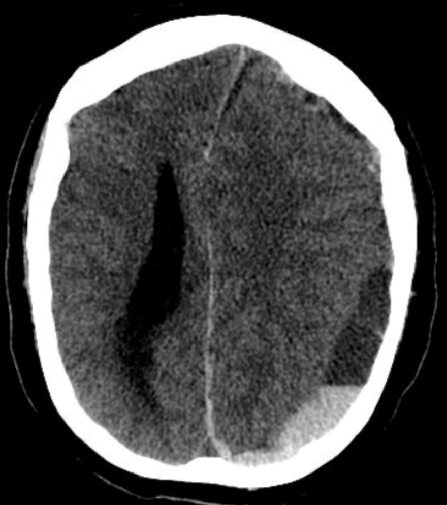
Which of the following is not true about non-contrast CT scan in head injury?
Which one of the following is a secondary brain injury?
Neurological status is assessed under which step of ABCDE of trauma care?
A man presents to the emergency department with a head injury following a vehicular accident. What is the investigation of choice?
A polytrauma patient's CT brain shows a crescent-shaped extra-axial collection with a concave inner margin. What is the most likely diagnosis?

A child with moderate to severe head injury is admitted in PICU. First line treatments are all except:
Explanation: ***Airway maintenance*** - Maintaining a **patent airway** is the absolute first step in managing any critically ill patient, including those with raised ICP, to ensure adequate **oxygenation and ventilation** [1]. - Without proper airway management, the brain will suffer from **hypoxia**, which can worsen cerebral edema and further increase ICP, leading to a poorer prognosis. *Breathing* - While essential in the **ABCs (Airway, Breathing, Circulation)**, ensuring adequate breathing (ventilation) comes immediately after securing the airway [1]. - An obstructed airway will prevent effective breathing, making airway maintenance the priority. *mannitol* - **Mannitol** is an osmotic diuretic used to reduce ICP by drawing fluid from the brain into the vasculature, but it is a **pharmacological intervention** that follows initial stabilization of the ABCs. - Administering mannitol without first securing the airway and ensuring ventilation could be detrimental if the patient is hypoxic. *Loading dose of phenytoin* - Administering a **loading dose of phenytoin** is primarily for seizure prophylaxis or treatment, which may be necessary in some cases of elevated ICP, but it is not the **immediate first step** in managing acute ICP elevation. - Seizure control is important, but airway, breathing, and circulation take precedence in the initial stabilization phase.
Explanation: ***20% Mannitol*** - **Mannitol** is an osmotic diuretic that reduces **intracranial pressure (ICP)** by creating an osmotic gradient, drawing water from the brain parenchyma into the intravascular space [1]. - Its rapid onset of action and significant ICP-reducing effects make it the drug of choice for acute management of elevated ICP in head injuries without hematoma. *Lasix* - **Furosemide (Lasix)** is a loop diuretic that can reduce ICP by decreasing cerebrospinal fluid production and promoting diuresis. - However, its effects are generally slower and less potent than mannitol for acute, rapidly increasing ICP. *Glycine* - **Glycine** is an amino acid and neurotransmitter; it has no direct role in the acute management of increased ICP. - It is sometimes used as an irrigating solution in urological procedures but is not indicated for brain injury. *Steroids* - **Steroids**, particularly **dexamethasone**, are effective in reducing vasogenic edema associated with brain tumors or abscesses. - They are generally **not recommended** for acute traumatic brain injury due to lack of benefit and potential for increased mortality or complications.
Explanation: ***Tachycardia*** - **Tachycardia** is generally *not* a sign of increased intracranial pressure (ICP); rather, **bradycardia** (Cushing's reflex) is a classic finding. - While other systemic responses may occur, a direct, consistent increase in heart rate due to elevated ICP is uncommon. *Papilledema* - **Papilledema** is a swelling of the **optic disc** due to increased ICP, a critical diagnostic sign [1]. - The increased pressure impedes venous return from the retina, causing the optic nerve head to bulge. *Headache* - **Headache** is a common and often early symptom of increased ICP due to the stretching of pain-sensitive meningeal and vascular structures [1]. - It is typically described as a dull, throbbing pain, often worse in the morning or with straining. *Seizures* - **Seizures** can result from increased ICP as the pressure on brain tissue can lead to electrical instability and abnormal neuronal discharge [2]. - This symptom indicates significant cortical irritation or dysfunction caused by the elevated pressure.
Explanation: ***Raccoon eyes seen in subgaleal hemorrhage*** - **Raccoon eyes** (periorbital ecchymosis) are typically seen with **anterior cranial fossa fractures**, not subgaleal hemorrhage. - Subgaleal hemorrhage is a collection of blood between the galea aponeurotica and the periosteum, usually causing diffuse **scalp swelling**. *Depressed skull is associated with brain injury at the immediate area of impact* - A depressed skull fracture means a portion of the skull is pushed inward, directly impacting the underlying **brain tissue**. - This can lead to localized **contusions**, **lacerations**, or **hematomas** at the site of impact. *Carotid-cavernous fistula occur in base skull* - **Carotid-cavernous fistulas** (CCF) commonly result from **traumatic rupture** of the internal carotid artery within the **cavernous sinus**. - This type of injury is often associated with **severe skull base fractures**, particularly those involving the sphenoid bone. *Post traumatic epilepsy seen in 15%* - The incidence of **post-traumatic epilepsy** (PTE) after severe head injury ranges from 5% to 15%, making 15% a plausible, though upper-end, estimate. - Risk factors for PTE include **depressed skull fractures**, **intracranial hematomas**, and **early seizures**.
Explanation: ***Subdural hematoma increases in density on serial CT scans over weeks*** - A **subdural hematoma (SDH)** typically **decreases in density** over weeks due to clot lysis and absorption of its proteinaceous components, transitioning from hyperdense (acute) to isodense (subacute) and then hypodense (chronic). - An increase in density on serial CT scans would imply continued bleeding or re-hemorrhage, which is not the typical natural progression of an acute SDH. *Extradural hematomas are usually lens-shaped* - **Extradural hematomas (EDH)** are typically **biconvex** or **lens-shaped** because they accumulate in the epidural space and are limited by cranial sutures where the dura is tightly adhered to the skull. - This characteristic shape helps distinguish them from subdural hematomas. *Acute subdural hematoma appears as crescent shadow of increased density* - An **acute subdural hematoma (SDH)** appears as a **crescent or crescent-shaped shadow** of increased density (hyperdense) on a non-contrast CT scan because it spreads along the inner surface of the dura, conforming to the brain's convexity. - This indicates active bleeding that is relatively fresh, usually within the first 3-7 days. *Subarachnoid hemorrhage appears as areas of increased density in basilar cisterns* - **Subarachnoid hemorrhage (SAH)** manifests as areas of **increased density (hyperdensity)** within the **basilar cisterns**, Sylvian fissures, or sulci, due to blood filling these cerebrospinal fluid (CSF)-containing spaces. - This finding is a key indicator of SAH on non-contrast CT.
Explanation: ***Intracerebral haematoma with raised intracranial pressure*** - **Intracerebral haematoma** is a potentially treatable, secondary injury directly contributing to **raised intracranial pressure (ICP)**, leading to further brain damage if not managed. - **Secondary brain injuries** occur minutes to days after the initial impact, resulting from a cascade of events like ischaemia, oedema, and intracranial hypertension. *Diffuse axonal injury* - **Diffuse axonal injury (DAI)** is a **primary brain injury** caused by shearing forces at the moment of impact. - It is a direct consequence of the initial trauma, not a subsequent physiological process. *Cortical lacerations* - **Cortical lacerations** are **primary injuries**, representing a direct tearing or cutting of brain tissue due to the initial traumatic force. - These are immediately present at the time of injury and are not a consequence of subsequent physiological changes. *Brainstem and hemispheric contusions* - **Contusions** are localised areas of bruising on the brain, characteristic of a **primary brain injury**, occurring directly from the impact. - While contusions can evolve and contribute to secondary injury mechanisms like oedema, the contusion itself is a direct result of the initial trauma.
Explanation: ***D - Disability: neurological status*** - The "D" in ABCDE trauma assessment specifically stands for **Disability**, which involves a rapid assessment of the patient's **neurological status**. - This step typically includes evaluating **level of consciousness** using tools like the AVPU scale (Alert, Voice, Pain, Unresponsive) or the Glasgow Coma Scale (GCS), assessing pupillary response, and identifying any gross motor deficits. *C - Circulation with haemorrhage control* - This step focuses on assessing and managing **blood flow**, including evaluating heart rate, blood pressure, capillary refill, and controlling any sources of external hemorrhage. - While neurological issues can result from poor circulation, the primary assessment of the nervous system itself is not performed here. *E - Exposure: completely undress the patient and assess for other injuries* - This final step involves a thorough **inspection of the entire body** to identify hidden injuries, such as bruising, lacerations, or deformities, while simultaneously ensuring temperature regulation. - It is for overall physical assessment, not for initial neurological evaluation. *B - Breathing and ventilation* - This step involves assessing the patient's **respiratory effort**, checking for symmetrical chest rise, listening to breath sounds, and intervening to ensure adequate oxygenation and ventilation. - While critical for brain function, this step focuses on the respiratory system, not the direct assessment of neurological function.
Explanation: ***NCCT*** - **Non-contrast Computed Tomography (NCCT)** of the head is the **investigation of choice** for acute head trauma due to its rapid acquisition, wide availability, and excellent sensitivity for detecting acute hemorrhage, fractures, and mass effects. - It rapidly identifies life-threatening conditions such as **epidural, subdural, and intracerebral hemorrhages**, which require immediate intervention. *MRI* - **MRI** is superior for detecting subtle brain tissue injuries, diffuse axonal injury, and non-hemorrhagic lesions but is generally **not the first-line investigation** in acute trauma due to longer scan times, limited availability in the emergency setting, and inability to detect acute hemorrhage as clearly as CT. - Its use is typically reserved for follow-up studies or when CT findings are inconclusive or specific soft tissue detail is required. *CECT* - **Contrast-enhanced CT (CECT)** of the head is reserved for specific indications like evaluating vascular lesions (e.g., aneurysms, arteriovenous malformations) or tumors, which are generally **not the primary concern** in the initial assessment of acute head trauma. - Administering contrast agents can delay imaging, may pose risks to patients with renal impairment or allergies, and does not significantly improve the detection of acute traumatic hemorrhage compared to NCCT. *X-ray* - **X-rays** of the skull are useful for detecting **skull fractures**, but they provide **limited information** regarding intracranial injuries or soft tissue damage, which are critical in head trauma. - They have largely been superseded by CT scans, which offer a more comprehensive view of both bony structures and intracranial contents.
Explanation: ***SDH*** - The image shows a **crescent-shaped collection** of hemorrhage with a concave inner margin, consistent with a **subdural hematoma** (SDH). - SDHs result from the tearing of **bridging veins** and typically conform to the brain's surface, crossing suture lines but not limited by bony sutures. *EDH* - An **epidural hematoma (EDH)** characteristically appears as a **lenticular** or **biconvex** shape on CT, not crescent-shaped. - EDHs are typically caused by arterial bleeding, often from the **middle meningeal artery**, and are limited by cranial sutures. *Contusion* - A **contusion** is brain tissue bruising that appears as **heterogeneous areas** of hemorrhage and edema within the brain parenchyma itself. - It would not manifest as a distinct extra-axial collection with a smooth, concave margin. *Diffuse axonal injury* - **Diffuse axonal injury (DAI)** involves widespread microscopic damage to axons, often at the gray-white matter junction. - It may appear as *punctate hemorrhages* or **small lesions** at these junctions on CT, but often the CT can be normal, and it would not present as a large extra-axial collection.
Explanation: ***IV mannitol*** - While **intravenous mannitol** is used in the management of head injury to reduce **intracranial pressure (ICP)**, it is **not a first-line treatment**. - It is a **second-line therapy** reserved for documented or suspected elevated ICP despite initial supportive measures. - First-line management focuses on maintaining adequate oxygenation, ventilation, and cerebral perfusion, while mannitol is used for specific ICP management when needed. *Analgesia and sedation* - **Analgesia and sedation** are essential **first-line treatments** to reduce pain, anxiety, and agitation, which can increase **intracranial pressure (ICP)**. - These therapies ensure patient comfort, decrease metabolic demand, facilitate mechanical ventilation, and prevent secondary brain injury. *Hypothermia* - **Therapeutic hypothermia** is **NOT routinely recommended** as a first-line treatment in pediatric traumatic brain injury. - Current evidence (including the Cool Kids trial) has not demonstrated benefit, and it may be associated with adverse effects. - It is considered **investigational** and not part of standard first-line management protocols. - **Note**: While this is also not first-line, the question specifically tests knowledge that mannitol is second-line therapy for ICP management. *Controlled mechanical ventilation* - **Controlled mechanical ventilation** is a fundamental **first-line treatment** for severe head injury to secure the airway and ensure adequate oxygenation and ventilation. - Prevents secondary brain injury from **hypoxia** and **hypercapnia**, which can worsen outcomes. - Maintaining appropriate **PaCO2 levels** is critical to control cerebral blood flow and intracranial pressure.
Get full access to all questions, explanations, and performance tracking.
Start For Free