What is the formula for calculating GFR?
A child presents with proteinuria, gross hematuria, sensorineural deafness, and anterior lenticonus. What is the inheritance pattern of this condition?
A 4-month-old infant with a urinary tract infection (UTI) was treated for 14 days with IV cefotaxime. What is the next step in management?
A child presented with frothy urine, massive proteinuria, and edema. Urine examination revealed no RBCs, no WBCs, no casts, and no crystals. There is no prior episode of similar presentation. What is your diagnosis?
A 5-year-old child develops the sudden onset of bloody diarrhea, vomiting of blood, hematuria, and renal failure following a flulike gastrointestinal illness. The blood urea nitrogen (BUN) level is markedly increased, but fibrin degradation products and blood clotting times are within normal limits. A peripheral blood smear reveals poikilocytes, schistocytes, and a decrease in the number of platelets. No fever or neurologic symptoms are present. What is the best diagnosis for this patient?
Regarding Hemolytic Uremic Syndrome (HUS), all of the following are true except:
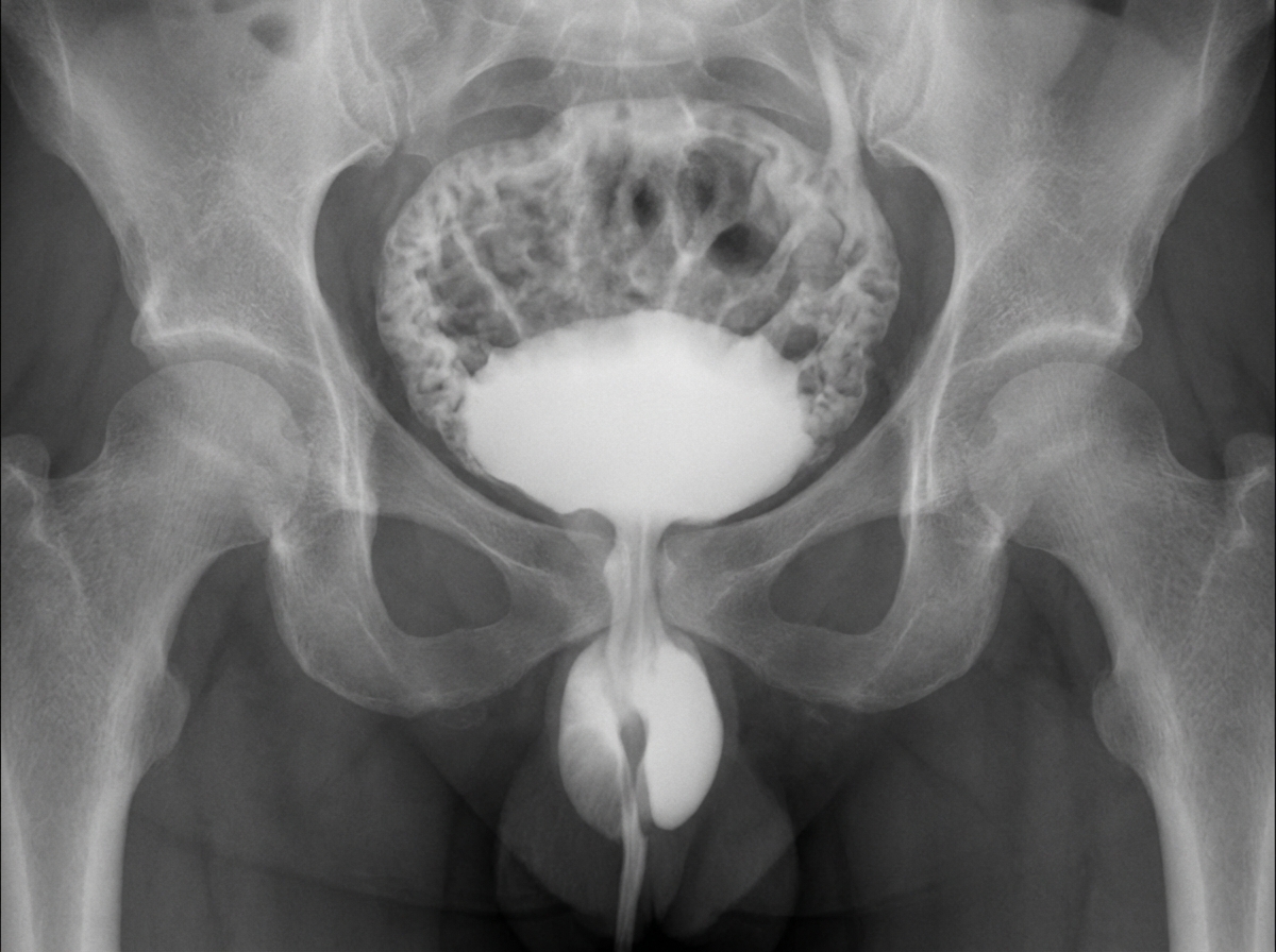
A male child presents with repeated urinary infections and failure to gain weight. A MCU was carried out as shown in the provided image. What is the most probable diagnosis?

Shigella associated hemolytic uremic syndrome is associated with all of the following except?
What is true about minimal change disease?
What is the marker for renal vasculitis in children?
Explanation: The correct answer is **Option A**. In pediatric medicine, the standard method for estimating the Glomerular Filtration Rate (eGFR) is the **Schwartz Formula**. ### **Why Option A is Correct** The Schwartz Formula is based on the physiological principle that creatinine production is proportional to muscle mass, which in children is directly related to their **height (length)**. * **Formula:** $eGFR = \frac{k \times \text{Height (cm)}}{\text{Serum Creatinine (mg/dL)}}$ * **The Constant (k):** This represents the creatinine excretion per unit of body size. It varies with age (e.g., 0.45 for infants, 0.413 in the updated "Bedside Schwartz" formula). ### **Why Other Options are Incorrect** * **Option B & C:** Weight is not used in the pediatric eGFR formula because weight fluctuates significantly with fluid status (edema) and nutrition, making it an unreliable surrogate for muscle mass compared to height. * **Option D:** This suggests an inverse relationship between height and GFR, which is mathematically incorrect. As a child grows taller and gains muscle mass, their creatinine production increases; therefore, height must be in the numerator to balance the equation. ### **High-Yield Clinical Pearls for NEET-PG** 1. **Bedside Schwartz Formula:** The most commonly used updated constant is **0.413**. It is used for children aged 1 to 16 years. 2. **Units:** Always remember height is measured in **centimeters (cm)** and serum creatinine in **mg/dL**. 3. **Cystatin C:** In children with abnormal muscle mass (e.g., neuromuscular wasting), Serum Cystatin C is a more accurate marker than creatinine as it is independent of muscle bulk. 4. **Gold Standard:** While Schwartz is used clinically, the gold standard for measuring GFR remains **Inulin clearance** (though rarely used in routine practice).
Explanation: **Explanation:** The clinical presentation of **proteinuria, gross hematuria, sensorineural deafness, and anterior lenticonus** is pathognomonic for **Alport Syndrome**. This condition is a hereditary type IV collagen disorder affecting the basement membranes of the kidney, cochlea, and eye. **Why "All of the above" is correct:** Alport Syndrome is genetically heterogeneous. While the most common form is **X-linked (85%)**, it can also be inherited through other patterns: 1. **X-linked Dominant (85%):** Due to mutations in the *COL4A5* gene. 2. **Autosomal Recessive (10-15%):** Due to mutations in *COL4A3* or *COL4A4* genes. 3. **Autosomal Dominant (Rare, ~5%):** Also due to mutations in *COL4A3* or *COL4A4* genes. Because the question asks for the inheritance pattern of "this condition" (Alport Syndrome) and all three patterns are documented in medical literature, "All of the above" is the most accurate choice. **Clinical Pearls for NEET-PG:** * **Pathogenesis:** Defect in **Type IV Collagen** (the "Main" collagen of basement membranes). * **Ocular Hallmark:** **Anterior Lenticonus** (conical protrusion of the lens) is virtually pathognomonic for Alport Syndrome. * **Electron Microscopy (High Yield):** Shows characteristic **"Basket-weave appearance"** due to irregular thinning and thickening of the Glomerular Basement Membrane (GBM). * **Hearing Loss:** Typically bilateral, progressive sensorineural hearing loss (SNHL) affecting high frequencies. * **Differential:** If a patient has hematuria but *normal* hearing and vision, consider **Thin Basement Membrane Disease** (Benign Familial Hematuria).
Explanation: ### Explanation The management of a first-time febrile Urinary Tract Infection (UTI) in infants and children has evolved to minimize invasive procedures. According to the **AAP (American Academy of Pediatrics)** and **Indian Society of Pediatric Nephrology (ISPN)** guidelines, the primary goal after the first UTI is to screen for structural abnormalities. **1. Why Option A is Correct:** **Ultrasound (USG) KUB** is the recommended first-line investigation for all infants (2 months to 2 years) after their first febrile UTI. It is non-invasive and effective at detecting structural anomalies like hydronephrosis, renal agenesis, or duplication. In this 4-month-old, USG is the immediate next step to screen for any gross anatomical defects. **2. Why Other Options are Incorrect:** * **Option B (USG + MCU):** A Micturating Cystourethrogram (MCU/VCUG) is no longer routine after the *first* UTI unless the USG shows hydronephrosis, scarring, or other suspicious findings. It is also indicated if the UTI is "atypical" (e.g., non-E. coli organism, septicemia) or "recurrent." * **Option C & D (DMSA):** A DMSA scan is used to detect acute pyelonephritis or permanent renal scarring. While highly sensitive, it is not a routine immediate investigation after a first UTI unless the clinical course is complicated or the USG is abnormal. **Clinical Pearls for NEET-PG:** * **MCU Timing:** If indicated, MCU should be performed after the infection has subsided (usually 2–4 weeks later) to avoid false positives due to infection-induced reflux. * **DMSA Timing:** To detect permanent scarring, DMSA should be performed **6 months** after the acute infection. * **Prophylaxis:** Routine antibiotic prophylaxis is generally not recommended after the first UTI unless high-grade Vesicoureteral Reflux (VUR) is diagnosed. * **Gold Standard for VUR:** MCU remains the gold standard for diagnosing and grading VUR.
Explanation: ### Explanation The clinical presentation of **frothy urine** (indicating significant proteinuria), **massive proteinuria**, and **edema** defines **Nephrotic Syndrome**. **1. Why Minimal Change Disease (MCD) is correct:** MCD is the most common cause of nephrotic syndrome in children (approx. 80% of cases). The hallmark of MCD is **"pure" nephrotic syndrome**, meaning it presents with massive proteinuria but lacks features of "nephritic" involvement. The absence of RBCs (hematuria), WBCs, and casts in the urine sediment is highly characteristic. In MCD, light microscopy shows normal glomeruli, but electron microscopy reveals **effacement of podocyte foot processes**. **2. Why other options are incorrect:** * **IgA Nephropathy:** This is a **nephritic** syndrome. It typically presents with recurrent episodes of gross or microscopic hematuria (RBCs in urine), often following an upper respiratory tract infection. * **Membranous Glomerulonephritis (MGN):** While it causes nephrotic syndrome, it is rare in children and more common in adults. It is often associated with secondary causes like Hepatitis B or SLE. * **Membranoproliferative Glomerulonephritis (MPGN):** This typically presents with a **mixed nephrotic-nephritic picture**, meaning urine would likely show hematuria and casts, along with low complement (C3) levels. **Clinical Pearls for NEET-PG:** * **Most common cause** of Nephrotic Syndrome in children: MCD. * **Selective Proteinuria:** MCD is characterized by the loss of albumin only (due to loss of polyanionic charge on the basement membrane). * **Treatment of Choice:** Corticosteroids (Prednisolone) are the first-line therapy; MCD is highly steroid-responsive. * **Diagnosis:** In children, a renal biopsy is usually **not** required initially if the presentation is classic; it is reserved for steroid-resistant cases.
Explanation: ### Explanation The clinical presentation describes the classic triad of **Hemolytic-Uremic Syndrome (HUS)**: Microangiopathic hemolytic anemia (MAHA), thrombocytopenia, and acute kidney injury (AKI). **1. Why HUS is the Correct Answer:** HUS typically follows a prodromal gastrointestinal illness (often caused by Shiga toxin-producing *E. coli* O157:H7). The toxin causes endothelial damage in the renal microvasculature, leading to platelet activation and microthrombi formation. As RBCs pass through these partially occluded vessels, they are mechanically shredded, resulting in **schistocytes** (fragmented cells) and **poikilocytes**. The consumption of platelets leads to thrombocytopenia, while the localized thrombi in the kidney cause renal failure (elevated BUN/Creatinine) and hematuria. **2. Why the Other Options are Incorrect:** * **DIC:** While DIC also features schistocytes and low platelets, it is characterized by **abnormal coagulation profiles** (prolonged PT/aPTT) and elevated Fibrin Degradation Products (FDPs). In this case, clotting times and FDPs are normal, ruling out DIC. * **TTP:** TTP shares the features of HUS but is more common in adults and typically presents with a **pentad** that includes fever and prominent **neurologic symptoms**, both of which are absent here. * **ITP:** ITP presents with isolated thrombocytopenia. It does not cause schistocytes, hemolytic anemia, or renal failure. **3. NEET-PG High-Yield Pearls:** * **Most common cause of AKI in children:** HUS. * **Classic Triad:** Hemolytic anemia (Coombs negative) + Thrombocytopenia + Renal failure. * **Key Lab Finding:** Normal PT/aPTT (distinguishes HUS from DIC). * **Management:** Primarily supportive (fluid/electrolyte balance, dialysis if needed). **Antibiotics and anti-motility agents are contraindicated** as they may increase toxin release and worsen the condition.
Explanation: **Explanation:** Hemolytic Uremic Syndrome (HUS) is a clinical triad of **Microangiopathic Hemolytic Anemia (MAHA)**, **Thrombocytopenia**, and **Acute Kidney Injury**. **Why Option B is the Correct Answer (The False Statement):** The hallmark of HUS is **Coombs-negative** hemolytic anemia. The hemolysis in HUS is mechanical, caused by the shearing of red blood cells as they pass through microthrombi in small vessels (fragmentation hemolysis), resulting in **schistocytes** on a peripheral smear. A positive Coombs test would instead suggest an autoimmune etiology, which is not the mechanism here. **Analysis of Other Options:** * **Option A:** In many Asian countries, including India, HUS is frequently associated with *Shigella dysenteriae* type 1 (producing Shiga toxin) rather than the *E. coli* O157:H7 (Verocytotoxin) strains more common in Western nations. * **Option C:** Typical D+ (Diarrhea-associated) HUS usually occurs as a single episode, and recurrences are rare. Recurrent cases often point toward "Atypical HUS" (complement-mediated). * **Option D:** Thrombocytopenia is a core feature due to platelet consumption in microthrombi. It is often transient, improving as the acute phase of the illness resolves. **Clinical Pearls for NEET-PG:** * **Triad:** Hemolytic anemia (Schistocytes), Thrombocytopenia, and Renal failure. * **Most common cause (Global):** Shiga-toxin producing *E. coli* (STEC/VTEC) O157:H7. * **Most common cause (India):** *Shigella dysenteriae* type 1. * **Management:** Primarily supportive (fluid/electrolyte balance, dialysis). **Antibiotics and anti-motility agents are contraindicated** in STEC-HUS as they may increase toxin release. * **Atypical HUS:** Associated with mutations in Factor H, Factor I, or Membrane Cofactor Protein (MCP).
Explanation: ***Posterior urethral valve*** - **Male child** with **recurrent UTIs** and **failure to thrive** along with characteristic **MCU findings** showing **dilated posterior urethra** and **trabeculated bladder** strongly suggests posterior urethral valve. - **PUV** causes **bladder outlet obstruction** leading to **vesicoureteral reflux**, **hydronephrosis**, and the classic **keyhole sign** on imaging studies. *Meatal stenosis* - Typically presents with **narrow urinary stream** and **dysuria** but does not cause the severe **bladder trabeculation** and **upper tract dilatation** seen on MCU. - Would show **narrowing at the urethral meatus** rather than **posterior urethral dilatation** characteristic of this case. *Bladder diverticulum* - Presents as **outpouching of bladder wall** on imaging and may cause **urinary stasis** but is uncommon in children. - Does not typically cause the **posterior urethral dilatation** and **keyhole appearance** seen in posterior urethral valves. *Bladder polyp* - Usually presents as **filling defects** within the bladder on imaging rather than **urethral dilatation**. - **Benign bladder masses** are rare in children and do not cause the characteristic **obstructive uropathy** pattern seen in this case.
Explanation: **Explanation:** Hemolytic Uremic Syndrome (HUS) is a clinical triad characterized by **Microangiopathic Hemolytic Anemia (MAHA)**, **Thrombocytopenia**, and **Acute Kidney Injury (AKI)**. In the context of *Shigella dysenteriae* type 1, the Shiga toxin causes endothelial damage, primarily in the renal vasculature. **Why Hyperkalemia is the correct answer (the "Except"):** While AKI typically leads to hyperkalemia due to decreased potassium excretion, Shigella-associated HUS is a unique exception. *Shigella* infection often causes severe **secretory diarrhea**, leading to significant gastrointestinal loss of potassium. Consequently, patients frequently present with **hypokalemia** or normal potassium levels, despite the presence of renal failure. **Analysis of Incorrect Options:** * **Thrombocytopenia:** This is a hallmark of HUS. Platelets are consumed during the formation of microthrombi in damaged small blood vessels. * **Neurological symptoms:** These occur in approximately 25–50% of cases due to metabolic encephalopathy or microvascular thrombi in the CNS, presenting as irritability, seizures, or altered consciousness. * **Renal microthrombi:** The pathophysiology involves Shiga toxin-induced endothelial injury, leading to the deposition of fibrin-platelet thrombi within the glomerular capillaries, which causes the characteristic renal failure. **High-Yield Clinical Pearls for NEET-PG:** * **Classic Triad:** Anemia (Coombs negative), Thrombocytopenia, and Renal failure. * **Peripheral Smear:** Will show **Schistocytes** (fragmented RBCs) and helmet cells. * **D+ HUS vs. D- HUS:** D+ (Diarrhea-associated) is most commonly caused by *E. coli* O157:H7 (Verotoxin) or *Shigella* (Shiga toxin). * **Management:** Primarily supportive (fluid/electrolyte balance, dialysis). **Antibiotics and anti-motility agents are generally avoided** as they may worsen toxin release.
Explanation: **Explanation:** Minimal Change Disease (MCD) is the most common cause of Nephrotic Syndrome in children, accounting for approximately 80-90% of cases in the pediatric age group. **1. Why Option A is correct:** The peak incidence of MCD occurs in early childhood, specifically between **2 to 4 years of age**. It is more common in boys than girls (2:1 ratio). In this age group, the presence of nephrotic syndrome is so strongly associated with MCD that a renal biopsy is usually deferred, and a trial of steroids is initiated immediately. **2. Why the other options are incorrect:** * **Option B:** Renal failure is **rare** in MCD. The prognosis is generally excellent, and most children maintain normal renal function. If a patient presents with persistent hypertension or azotemia, alternative diagnoses like Focal Segmental Glomerulosclerosis (FSGS) should be considered. * **Option C:** Steroids (Prednisolone) are the **mainstay of treatment**. Over 90% of children with MCD are "Steroid Sensitive," meaning they achieve complete remission within 4-8 weeks of therapy. * **Option D:** While MCD was traditionally associated with **selective proteinuria** (primarily albuminuria due to loss of the glomerular basement membrane's negative charge), this is no longer considered a pathognomonic or reliable diagnostic feature in modern clinical practice. The most definitive "true" epidemiological fact among the choices is the age of incidence. **NEET-PG High-Yield Pearls:** * **Light Microscopy:** Glomeruli appear normal (hence "Minimal Change"). * **Electron Microscopy (Gold Standard):** Shows **effacement (fusion) of podocyte foot processes**. * **Immunofluorescence:** Typically negative for immune deposits. * **Association:** Can be associated with Hodgkin’s Lymphoma in adults. * **Most common complication:** Infections (due to loss of IgG and complement factors in urine).
Explanation: **Explanation:** The question refers to **Henoch-Schönlein Purpura (HSP)**, now commonly known as **IgA Vasculitis**. It is the most common systemic vasculitis in children and frequently involves the kidneys (HSP Nephritis). **Why Option A is Correct:** The hallmark of HSP is the systemic deposition of **IgA-dominant immune complexes** in small vessels. In the kidneys, these complexes deposit in the mesangium, leading to glomerulonephritis. Approximately **50-70% of children** with HSP demonstrate **elevated serum IgA levels** during the acute phase. While not 100% sensitive, it serves as the primary laboratory marker associated with the pathogenesis of this specific pediatric vasculitis. **Analysis of Incorrect Options:** * **B. Low complement level:** HSP is a **normocomplementemic** condition. Low complement levels (C3, C4) are characteristic of Post-Streptococcal Glomerulonephritis (PSGN), Lupus Nephritis, or Membranoproliferative GN. * **C. Increased ANCA titre:** ANCA is the marker for small-vessel vasculitides like Granulomatosis with Polyangiitis (Wegener's) or Microscopic Polyangiitis, which are rare in the pediatric population compared to HSP. * **D. Increased ANA:** This is the screening marker for Systemic Lupus Erythematosus (SLE), not a primary marker for pediatric renal vasculitis. **High-Yield Clinical Pearls for NEET-PG:** * **Classic Tetrad of HSP:** Non-thrombocytopenic palpable purpura (lower limbs/buttocks), arthralgia, abdominal pain (intussusception risk), and renal involvement. * **Renal Biopsy:** Shows mesangial IgA deposits (identical to IgA Nephropathy/Berger’s disease). * **Prognosis:** Most cases are self-limiting; however, the long-term prognosis depends entirely on the severity of **renal involvement**.
Urinary Tract Infections
Practice Questions
Vesicoureteral Reflux
Practice Questions
Glomerulonephritis
Practice Questions
Nephrotic Syndrome
Practice Questions
Acute Kidney Injury
Practice Questions
Chronic Kidney Disease
Practice Questions
Renal Tubular Disorders
Practice Questions
Congenital Anomalies of the Kidney
Practice Questions
Hydronephrosis
Practice Questions
Hypertension in Children
Practice Questions
Hemolytic Uremic Syndrome
Practice Questions
Renal Replacement Therapy in Children
Practice Questions
Get full access to all questions, explanations, and performance tracking.
Start For Free