
Neonatology — MCQs

On this page
Which of the following statements is FALSE regarding kernicterus?
What is the diagnosis in a baby girl delivered at 30 weeks' gestation?
Two days after birth, a child developed respiratory distress and had a scaphoid abdomen. Breath sounds were decreased on the left side. After bag and mask ventilation, an endotracheal tube was inserted, and the maximal cardiac impulse shifted to the right side. What should be the next step in management?
A neonate continuously regurgitates all feeds and is continuously drooling saliva. Which of the following is the most probable diagnosis?
What is the definition of hypertension in a full-term newborn?
About trisomy 13, which of the following is a true statement?
Which vitamin is administered to newborns to prevent hemorrhage?
What is the ponderal index in malnourished babies?
A premature baby born to a diabetic mother presented with respiratory distress and was admitted to the neonatal intensive care unit. Which of the following findings indicates Respiratory Distress Syndrome (RDS) in a newborn?
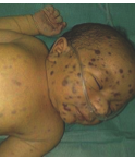
A boy presents with multiple bullous lesions on his trunk and periostitis observed on x-rays. What is the most appropriate next investigation?
Practice by Chapter
Neonatal Resuscitation
Practice Questions
Care of the Normal Newborn
Practice Questions
Prematurity and Low Birth Weight
Practice Questions
Respiratory Distress Syndrome
Practice Questions
Neonatal Jaundice
Practice Questions
Neonatal Sepsis
Practice Questions
Necrotizing Enterocolitis
Practice Questions
Intraventricular Hemorrhage
Practice Questions
Persistent Pulmonary Hypertension
Practice Questions
Perinatal Asphyxia
Practice Questions
Neonatal Seizures
Practice Questions
Congenital Anomalies
Practice Questions
Want unlimited practice?
Get full access to all questions, explanations, and performance tracking.
Scan to download app