
Neonatology — MCQs

On this page
All are true about necrotizing enterocolitis except?
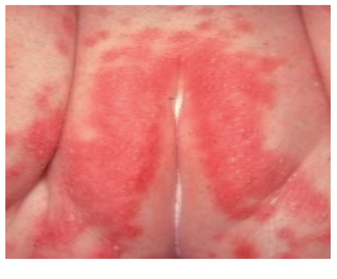
A rash is observed in a neonate admitted to the NICU for 6 days. What is the most likely cause?
Early onset sepsis in a neonate occurs within which timeframe?
Regarding Continuous Positive Airway Pressure (CPAP), all the following statements are true EXCEPT:
Which of the following features is NOT typically seen in the cold injury of neonates?
Gynecomastia in neonates is typically caused by which of the following?
A premature baby of 34 weeks gestation was delivered and subsequently developed bullous lesions on the skin. Radiographic examination revealed periostitis. What is the most appropriate next investigation?
A neonate presents with extensor posture at 8 days of age. What is the most likely diagnosis?
Cord cutting should be delayed in which condition?
Which of the following is an abnormal finding in a neonate?
Practice by Chapter
Neonatal Resuscitation
Practice Questions
Care of the Normal Newborn
Practice Questions
Prematurity and Low Birth Weight
Practice Questions
Respiratory Distress Syndrome
Practice Questions
Neonatal Jaundice
Practice Questions
Neonatal Sepsis
Practice Questions
Necrotizing Enterocolitis
Practice Questions
Intraventricular Hemorrhage
Practice Questions
Persistent Pulmonary Hypertension
Practice Questions
Perinatal Asphyxia
Practice Questions
Neonatal Seizures
Practice Questions
Congenital Anomalies
Practice Questions
Want unlimited practice?
Get full access to all questions, explanations, and performance tracking.
Scan to download app