
Neonatology — MCQs

On this page
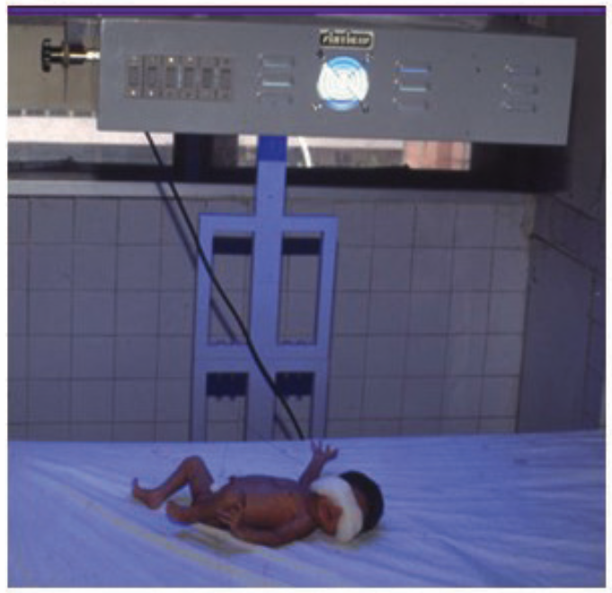
What wavelength is used in the given instrument to treat neonatal pathology?
All of the following are causes of bronchopulmonary dysplasia except?
What is the most common presentation of congenital Cytomegalovirus (CMV) infection?
During neonatal resuscitation of a spontaneously breathing preterm baby of 30 weeks gestation with labored breathing, oxygen delivery can be achieved by which of the following methods?
A very preterm baby on 30mL/kg of enteral feeding developed sudden severe abdominal distension with visible bowel loops on day 6 of life. The baby also showed temperature instability and lethargy. X-ray of the abdomen showed portal venous gas. What is the staging of NEC?
What is a sternocleidomastoid tumor?
Which of the following statements regarding late-onset Hemorrhagic Disease of the Newborn (HDN) is not true?
A 4-month-old boy is brought to the clinic. His parents report that the child stopped breathing at home, turned blue around his lips, and felt limp. After vigorous shaking of the infant and several mouth-to-mouth breaths, the boy's color returned to normal, and he resumed breathing. The infant's condition is best described as:
What is the recommended room temperature for maintaining warmth for neonates?
A newborn has a heart rate of 110, has been crying vigorously, with good muscle tone and active movements. The baby has good respiratory effort and rate. The body of the baby is pink in color but the extremities are blue. What is the APGAR score of the child?
Practice by Chapter
Neonatal Resuscitation
Practice Questions
Care of the Normal Newborn
Practice Questions
Prematurity and Low Birth Weight
Practice Questions
Respiratory Distress Syndrome
Practice Questions
Neonatal Jaundice
Practice Questions
Neonatal Sepsis
Practice Questions
Necrotizing Enterocolitis
Practice Questions
Intraventricular Hemorrhage
Practice Questions
Persistent Pulmonary Hypertension
Practice Questions
Perinatal Asphyxia
Practice Questions
Neonatal Seizures
Practice Questions
Congenital Anomalies
Practice Questions
Want unlimited practice?
Get full access to all questions, explanations, and performance tracking.
Scan to download app