
Neonatology — MCQs

On this page
A 7-day-old infant presents with symptoms of neonatal septicemia. What is the most likely cause?
A large-for-gestational age newborn infant was born 50 hours ago by cesarean section to a 26-year-old primigravida mother with insulin-dependent gestational diabetes. The infant's initial glucose was 25 mg/dL, but after feeding subsequent glucoses have all been above 60 mg/dL. The infant is now diaphoretic and irritable, and seems to have some twitching and tremors of the extremities. What is the most likely cause of this infant's problems?
The salmon patch (nevus simplex) typically disappears by what age?
What is a long-term complication of bronchopulmonary dysplasia?
Which of the following conditions in a newborn typically resolves spontaneously, EXCEPT?
A baby is said to be small for gestational age if its weight is below which percentile?
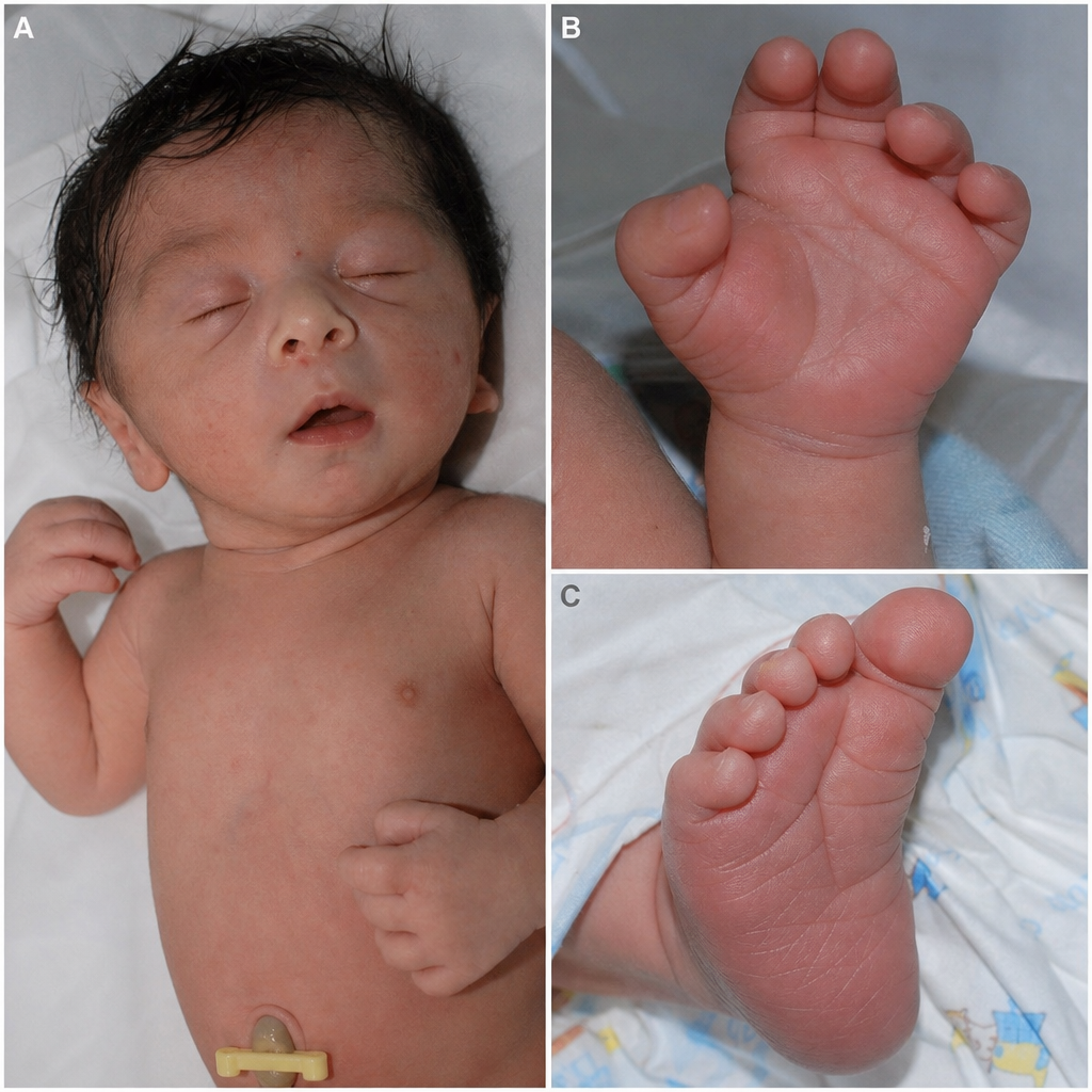
What is the condition affecting this newborn with facial dysmorphisms and the features shown below?
What are the FDA indications for inhaled nitric oxide?
Which of the following does not establish a diagnosis of congenital Cytomegalovirus (CMV) infection in a neonate?
Extremely low birth weight (ELBW) child is defined as?
Practice by Chapter
Neonatal Resuscitation
Practice Questions
Care of the Normal Newborn
Practice Questions
Prematurity and Low Birth Weight
Practice Questions
Respiratory Distress Syndrome
Practice Questions
Neonatal Jaundice
Practice Questions
Neonatal Sepsis
Practice Questions
Necrotizing Enterocolitis
Practice Questions
Intraventricular Hemorrhage
Practice Questions
Persistent Pulmonary Hypertension
Practice Questions
Perinatal Asphyxia
Practice Questions
Neonatal Seizures
Practice Questions
Congenital Anomalies
Practice Questions
Want unlimited practice?
Get full access to all questions, explanations, and performance tracking.
Scan to download app