
Neonatology — MCQs

On this page
The classic triad of congenital rubella syndrome includes which of the following, except?
A baby is born prematurely at 29 weeks gestation by caesarean delivery for fetal distress. The neonate develops tachypnea, nasal flaring, subcostal and intercostal retractions immediately after birth. Chest radiography shows bilateral, diffuse, ground glass appearance, air bronchograms, and poor lung expansion. What is the best treatment plan for this neonate?
A 28-week pregnant multigravida has an abnormal glucose tolerance test. Her previous child weighed 4 kg at birth. Which of the following is TRUE about infants of diabetic mothers?
Which of the following is true about erythema toxicum neonatorum?
A newborn infant presents with intermittent cyanosis that improves with crying and worsens when quiet. What is the most likely diagnosis?
When severe dehydration in a neonate occurs, what is the recommended fluid replacement amount in the first hour?
Which of the following is NOT a component of VACTERL association?
What is the birth weight of a baby considered to be low birth weight?
Which of the following is NOT associated with jaundice in infancy?
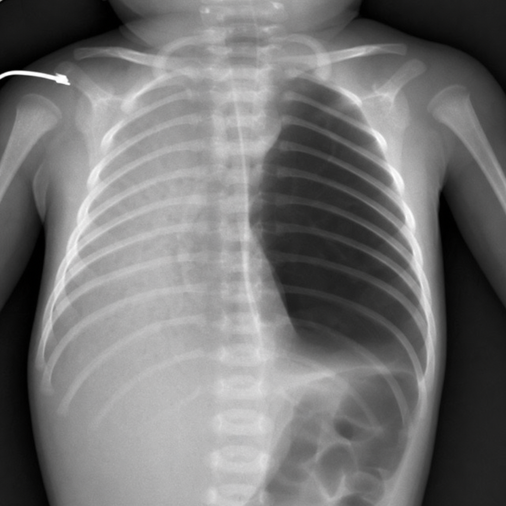
A neonate with respiratory distress and the given chest X-ray most likely has which of the following conditions?
Practice by Chapter
Neonatal Resuscitation
Practice Questions
Care of the Normal Newborn
Practice Questions
Prematurity and Low Birth Weight
Practice Questions
Respiratory Distress Syndrome
Practice Questions
Neonatal Jaundice
Practice Questions
Neonatal Sepsis
Practice Questions
Necrotizing Enterocolitis
Practice Questions
Intraventricular Hemorrhage
Practice Questions
Persistent Pulmonary Hypertension
Practice Questions
Perinatal Asphyxia
Practice Questions
Neonatal Seizures
Practice Questions
Congenital Anomalies
Practice Questions
Want unlimited practice?
Get full access to all questions, explanations, and performance tracking.
Scan to download app