What age group is most susceptible to sepsis?
All of the following decrease the risk of necrotizing enterocolitis (NEC) except?
Congenital long QT syndrome is associated with neonatal:
At what gestational age does the switchover from fetal to adult hemoglobin synthesis begin?
Maternal rubella infection during pregnancy can cause all of the following in the newborn EXCEPT:
A 5-day-old term male neonate presents with delayed passage of meconium, abdominal distension, and bilious vomiting. What is the diagnosis?
In neonatal cholestasis, what is the threshold for elevated direct bilirubin?
A newborn presents with respiratory distress with a respiratory rate of 86/min, nasal flaring, audible grunting, asynchronous chest and abdominal breathing, and no lower chest or xiphoid retractions. What is the Silverman score?
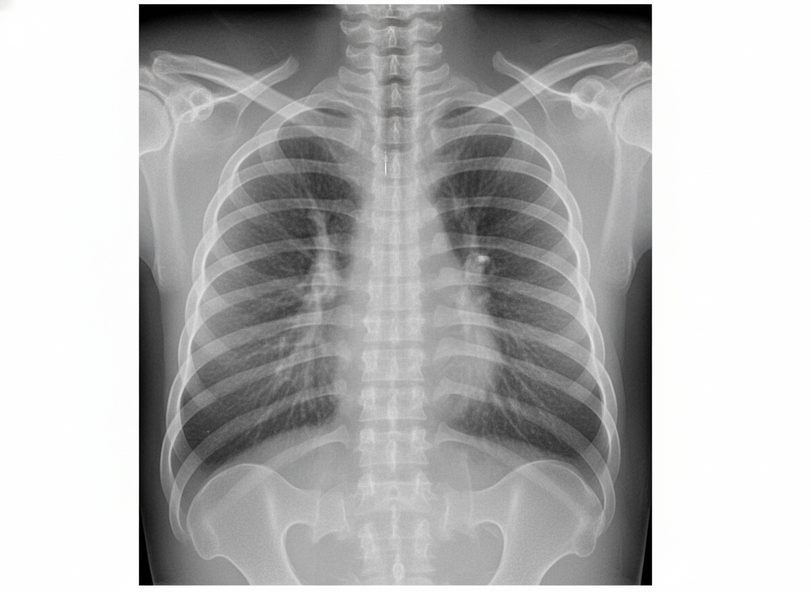
A term neonate presents with mild respiratory distress soon after birth. Chest X-ray shows typical findings. How is this condition managed?

What is the most common organism causing neonatal meningitis?
Explanation: **Explanation:** The susceptibility to sepsis follows a **U-shaped distribution**, with the highest incidence occurring at the extremes of age: neonates and the elderly. However, statistically and clinically, the **elderly (age >65 years)** represent the most susceptible group. **1. Why the Elderly are most susceptible:** The primary mechanism is **immunosenescence**—the progressive decline in immune function associated with aging. This includes decreased T-cell diversity, impaired phagocytosis by neutrophils, and a blunted cytokine response. Additionally, the elderly often have multiple comorbidities (diabetes, COPD, heart failure), increased use of indwelling devices (catheters), and frequent hospitalizations, all of which significantly elevate the risk of overwhelming infection and subsequent organ dysfunction. **2. Analysis of Incorrect Options:** * **Infants (Option A):** While neonates (especially preterm) have immature immune systems and are highly vulnerable, the absolute volume of sepsis cases and the cumulative risk factors are higher in the geriatric population. In many datasets, the incidence rate in those >85 years is nearly 10 times higher than in younger cohorts. * **Adolescents & Young Adults (Options B & D):** These groups typically have the most robust innate and adaptive immune responses. Sepsis in these age groups is rare and usually associated with specific triggers like trauma, meningococcemia, or underlying primary immunodeficiency. **Clinical Pearls for NEET-PG:** * **Definition:** Sepsis is now defined (Sepsis-3) as life-threatening organ dysfunction caused by a dysregulated host response to infection. * **qSOFA Score:** A quick bedside tool (Respiratory rate ≥22/min, Altered mentation, Systolic BP ≤100 mmHg). * **Most common source:** The **lung (Pneumonia)** is the most common site of infection leading to sepsis in the elderly. * **Neonatal Sepsis:** Remember that *Group B Streptococcus* (GBS) is a leading cause globally, though *Gram-negative organisms* (like E. coli and Klebsiella) are more common in the Indian context.
Explanation: **Explanation:** Necrotizing Enterocolitis (NEC) is a devastating gastrointestinal emergency primarily affecting preterm infants. Its pathogenesis involves a triad of intestinal immaturity, formula feeding, and microbial dysbiosis. **Why Lipoprotein Lipase (Option D) is the correct answer:** Lipoprotein lipase (LPL) is an enzyme responsible for the hydrolysis of triglycerides into free fatty acids. It has **no established role** in reducing the risk of NEC. In fact, elevated levels of free fatty acids (products of LPL activity) can sometimes be cytotoxic to the intestinal mucosa. Therefore, it does not serve a protective function against NEC. **Analysis of Incorrect Options (Protective Factors):** * **Lactoferrin (Option A):** An iron-binding glycoprotein found in breast milk. It has antimicrobial, anti-inflammatory, and immunomodulatory properties that help maintain gut integrity and prevent pathogen overgrowth. * **Probiotics (Option B):** Supplementation with *Lactobacillus* and *Bifidobacterium* helps colonize the gut with beneficial flora, competitive inhibition of pathogens, and enhancement of the mucosal barrier. * **L-arginine (Option C):** Arginine is a precursor to **Nitric Oxide (NO)**, a potent vasodilator. Supplementation improves mucosal perfusion and prevents ischemic injury to the bowel wall. **NEET-PG High-Yield Pearls:** * **Most important risk factor:** Prematurity and low birth weight. * **Best preventive strategy:** Exclusive Breastfeeding (contains IgA, lactoferrin, and oligosaccharides). * **Pathognomonic X-ray finding:** Pneumatosis intestinalis (gas in the bowel wall). * **Modified Bell’s Staging:** Used for clinical grading and management. * **Antenatal Steroids:** Also significantly reduce the risk of NEC by accelerating gut maturation.
Explanation: **Explanation:** **1. Why Sinus Bradycardia is Correct:** Congenital Long QT Syndrome (LQTS) is a channelopathy characterized by delayed repolarization of the myocardium. In neonates, this delay in the electrical cycle often manifests as **fetal or neonatal sinus bradycardia**. The underlying mechanism involves a prolonged refractory period of the ventricular myocardium, which can exert an inhibitory effect on the sinoatrial (SA) node or result in a functional 2:1 atrioventricular (AV) block. Bradycardia is often the earliest clinical sign of LQTS and can be detected in utero during routine fetal monitoring. **2. Why Other Options are Incorrect:** * **Sinus Tachycardia:** This is physiologically opposite to the presentation of LQTS. LQTS patients typically have a lower-than-normal heart rate for their age. * **Supraventricular Tachycardia (SVT):** While SVT is a common neonatal arrhythmia, it is not specifically associated with the pathophysiology of LQTS. * **Ventricular Tachycardia (VT):** While LQTS can lead to a specific type of polymorphic VT known as *Torsades de Pointes*, the question asks for the most characteristic neonatal association. Sinus bradycardia is a more consistent finding in the neonatal period and often precedes the development of life-threatening ventricular arrhythmias. **3. High-Yield Clinical Pearls for NEET-PG:** * **Jervell and Lange-Nielsen Syndrome:** Autosomal recessive LQTS associated with **sensorineural deafness**. * **Romano-Ward Syndrome:** Autosomal dominant LQTS (more common, no deafness). * **Torsades de Pointes:** The classic "twisting of points" polymorphic VT seen in LQTS; treated with **Magnesium Sulfate**. * **Neonatal Screening:** Any neonate with persistent unexplained bradycardia should receive an ECG to measure the QTc interval (Normal <440ms; in neonates, >470ms is suspicious).
Explanation: ### Explanation **Underlying Medical Concept** Hemoglobin synthesis in humans undergoes a programmed "switching" process. Fetal hemoglobin (HbF, $\alpha_2\gamma_2$) is the predominant type during intrauterine life due to its high oxygen affinity. The transition from the synthesis of $\gamma$-globin (fetal) to $\beta$-globin (adult) is a developmentally regulated process that is **not** triggered by birth, but rather by the gestational age of the fetus. This switchover begins at approximately **36 weeks of gestation**. By birth, adult hemoglobin (HbA, $\alpha_2\beta_2$) accounts for about 20–30% of total hemoglobin, and the process continues postnatally until HbF levels drop to <1% by 6–12 months of age. **Analysis of Options** * **Option A (30 weeks):** At this stage, the fetus is still almost entirely dependent on HbF for efficient oxygen extraction from maternal blood. * **Option B (36 weeks):** **Correct.** This is the physiological timing when the $\beta$-globin gene expression significantly increases, marking the start of the transition to HbA. * **Option C & D (Postnatal):** These are incorrect because the switch is a pre-programmed genetic event that begins **in utero**. While the *predominance* of HbA occurs postnatally, the *switchover* starts before birth. **NEET-PG High-Yield Pearls** * **HbF Structure:** $\alpha_2\gamma_2$. It does not bind 2,3-BPG effectively, leading to a leftward shift in the oxygen dissociation curve (higher affinity). * **Site of Erythropoiesis:** Yolk sac (3–8 weeks) $\rightarrow$ Liver (6–30 weeks, primary site) $\rightarrow$ Bone Marrow (begins at 18–20 weeks; becomes primary site by 30 weeks). * **Clinical Correlation:** Conditions like $\beta$-Thalassemia major do not manifest at birth because HbF levels are still high; symptoms appear only after 6 months when the switch to HbA is nearly complete.
Explanation: **Explanation:** Congenital Rubella Syndrome (CRS) is caused by the rubella virus crossing the placenta, primarily during the first trimester. The virus is non-cytolytic but inhibits mitosis, leading to organ hypoplasia and characteristic malformations. **Why Neural Tube Defects (NTDs) is the correct answer:** Neural tube defects (like anencephaly or spina bifida) are primarily associated with **folate deficiency** during the periconceptional period or certain teratogens like valproate. Rubella infection does not interfere with neural tube closure; therefore, NTDs are not a feature of CRS. **Why the other options are incorrect (Features of CRS):** * **Cardiovascular defects:** These occur in about 50% of cases. The most common lesion is **Patent Ductus Arteriosus (PDA)**, followed by peripheral pulmonary artery stenosis. * **Cataract:** Ocular abnormalities are a hallmark. "Pearls-and-nuclear" cataracts and microphthalmia are common. Another high-yield finding is **"Salt and pepper" retinopathy**. * **Deafness:** Sensorineural hearing loss is the **most common** clinical manifestation of CRS and may be the only finding in late-gestation infections. **High-Yield Clinical Pearls for NEET-PG:** * **Gregg’s Triad:** The classic triad of CRS includes **Cataract, Cardiac defects (PDA), and Deafness**. * **Dermatology:** Look for the **"Blueberry muffin rash"** (extramedullary hematopoiesis), also seen in CMV. * **Radiology:** "Celery stalking" (longitudinal radiolucent striations) in the metaphysis of long bones. * **Timing:** The risk of malformation is highest (up to 80%) if the mother is infected within the first 12 weeks of pregnancy.
Explanation: **Explanation:** The clinical triad of **delayed passage of meconium** (>48 hours), **abdominal distension**, and **bilious vomiting** in a neonate is a classic presentation of **Congenital Aganglionic Megacolon (Hirschsprung Disease)**. **Why Option A is Correct:** Hirschsprung disease is caused by the failure of neural crest cells to migrate cranio-caudally, resulting in an **absence of ganglion cells** (Auerbach’s and Meissner’s plexuses) in the distal colon. This leads to a functional obstruction because the aganglionic segment cannot relax, causing proximal bowel dilatation. It is the most common cause of lower intestinal obstruction in neonates. **Why Other Options are Incorrect:** * **B. Hypertrophic Pyloric Stenosis:** Typically presents at **3–6 weeks** of age with **non-bilious**, projectile vomiting and a palpable olive-shaped mass. It does not cause delayed meconium. * **C. Cystic Fibrosis:** While it causes Meconium Ileus (another cause of delayed meconium), the question describes the classic presentation of Hirschsprung. In Meconium Ileus, the obstruction is due to inspissated secretions in the terminal ileum, not aganglionosis. * **D. Intestinal Malrotation:** Usually presents with acute onset bilious vomiting due to midgut volvulus, but it is not typically associated with a primary delay in passing meconium. **NEET-PG High-Yield Pearls:** * **Gold Standard Diagnosis:** Rectal suction biopsy (shows absence of ganglion cells and hypertrophied nerve bundles). * **Initial Screening:** Contrast enema (shows a "transition zone"). * **Physical Exam:** "Squirt sign" or "Blast sign" (explosive release of stool/gas on digital rectal exam). * **Association:** Strongly associated with **Down Syndrome** (Trisomy 21).
Explanation: **Explanation:** Neonatal cholestasis is defined as prolonged physiological jaundice or pathological jaundice characterized by an accumulation of conjugated bilirubin in the serum. According to the latest clinical guidelines (NASPGHAN/ESPGHAN), the biochemical threshold for diagnosing cholestasis is a **conjugated (direct) bilirubin level >1.0 mg/dL**, regardless of the total serum bilirubin (TSB) level. **Why Option C is correct:** Historically, and still frequently tested in exams like NEET-PG, the definition of conjugated hyperbilirubinemia was based on the ratio of direct to total bilirubin. If the **TSB is >5 mg/dL**, a direct bilirubin fraction of **>20% of the total** is considered the diagnostic threshold for cholestasis. This threshold is critical because it necessitates an immediate workup to rule out life-threatening conditions like Biliary Atresia. **Why other options are incorrect:** * **Options A, B, and D:** These percentages (10%, 15%, and 25%) do not align with the standardized diagnostic criteria used in pediatric hepatology. While some older texts mentioned 15%, the consensus for "elevated" direct bilirubin in the context of a high TSB remains >20%. **High-Yield Clinical Pearls for NEET-PG:** 1. **The "Rule of 1.0":** Modern guidelines emphasize that any direct bilirubin **>1.0 mg/dL** is abnormal, even if it represents less than 20% of the TSB. 2. **Biliary Atresia (BA):** The most common surgical cause of neonatal cholestasis. The "Gold Standard" for diagnosis is an **Intraoperative Cholangiogram (IOCG)**. 3. **Kasai Procedure:** For BA, surgery (Hepatoportoenterostomy) is most successful if performed before **60 days of life**. 4. **Stool Color:** Pale or "clay-colored" (acholic) stools are a hallmark clinical sign of cholestasis.
Explanation: The **Silverman-Anderson Score** is a clinical tool used to assess the severity of respiratory distress in neonates. Unlike the Apgar score, a **higher** Silverman score indicates **greater** respiratory distress. ### **Calculation for this Case:** The score evaluates five parameters, each graded from 0 to 2: 1. **Upper Chest Movement:** Asynchronous (See-saw) breathing = **2 points** (Synchronous = 0, Lag = 1). 2. **Lower Chest Retractions:** None = **0 points** (Mild = 1, Marked = 2). 3. **Xiphoid Retractions:** None = **0 points** (Mild = 1, Marked = 2). 4. **Nasal Flaring:** Present (assumed mild/moderate) = **1 point** (None = 0, Marked = 2). 5. **Expiratory Grunting:** Audible with naked ear = **2 points** (None = 0, Audible with stethoscope = 1). **Total Score: 2 (Chest) + 0 (Lower) + 0 (Xiphoid) + 1 (Flaring) + 2 (Grunt) = 5.** ### **Analysis of Options:** * **A (1) & B (3):** These underestimate the severity. The presence of audible grunting and see-saw breathing alone contributes 4 points. * **D (6):** This overestimates the score. Since there are no lower chest or xiphoid retractions, the score cannot reach 6. ### **NEET-PG High-Yield Pearls:** * **Interpretation:** 0 = No distress; 1–3 = Mild; 4–6 = Moderate; >6 = Severe; 10 = Impending respiratory failure. * **Downe’s Score:** Used for both term and preterm babies (includes Cyanosis and Air entry), whereas Silverman is specifically preferred for **preterm** neonates. * **Mnemonic:** Remember **"Upper, Lower, Xiphoid, Nasal, Grunt"** to recall the five parameters in order.
Explanation: ***Supportive care as it resolves spontaneously*** - **Transient Tachypnea of the Newborn (TTN)** is caused by **delayed clearance of fetal lung fluid** and typically resolves within 24-48 hours without specific intervention. - Management involves **observation**, **oxygen supplementation** if needed, and ensuring adequate **fluid intake** while avoiding overhydration. *Oxygen delivery by CPAP* - **CPAP** is primarily indicated for **Respiratory Distress Syndrome (RDS)** or other conditions requiring **positive pressure ventilation**. - TTN rarely requires CPAP as it responds well to **minimal oxygen support** and **conservative management**. *Surfactant provided by InSURre technique* - **Surfactant therapy** is specifically indicated for **RDS** caused by **surfactant deficiency**, typically in preterm infants. - TTN involves **fluid retention** rather than surfactant deficiency, making surfactant therapy **ineffective** for this condition. *Oxygen delivery by endotracheal intubation* - **Endotracheal intubation** is reserved for severe respiratory failure requiring **mechanical ventilation** support. - TTN presents with **mild respiratory distress** and rarely progresses to require invasive ventilatory support.
Explanation: **Explanation:** Neonatal meningitis is a critical condition with high morbidity and mortality. In the neonatal period (0–28 days), the most common causative organisms are those found in the maternal birth canal. **Why E. coli is correct:** While **Group B Streptococcus (GBS)** is the leading cause of neonatal meningitis globally (especially in Western countries), **Gram-negative bacilli**, specifically **Escherichia coli**, are the most common cause in the **Indian subcontinent**. E. coli (particularly the K1 capsular strain) is the most frequent isolate in many Indian neonatal intensive care units, followed closely by Klebsiella. **Analysis of Incorrect Options:** * **B. Listeria monocytogenes:** While a classic cause of neonatal sepsis and meningitis (often associated with unpasteurized dairy), it is significantly less common than E. coli and GBS. * **C. Pseudomonas:** This is typically an opportunistic pathogen. It is a common cause of **nosocomial (hospital-acquired)** late-onset sepsis in NICUs but is not the most common cause of neonatal meningitis overall. * **D. Staph aureus:** This organism more commonly causes skin, soft tissue, and bone infections (osteomyelitis). While it can cause late-onset sepsis, it is a rare primary cause of meningitis. **High-Yield Clinical Pearls for NEET-PG:** 1. **Global vs. India:** If the question asks for the most common cause worldwide, the answer is **Group B Streptococcus (S. agalactiae)**. In the Indian context, prioritize **E. coli**. 2. **Late-onset Meningitis:** Beyond the first week of life, *Coagulase-negative Staphylococci* (CONS) and *Klebsiella* become more prevalent. 3. **Empiric Treatment:** The standard initial antibiotic regimen is typically **Ampicillin + Gentamicin** (or a third-generation cephalosporin like Cefotaxime) to cover both GBS and Gram-negative enteric bacilli. 4. **Clinical Sign:** A **bulging fontanelle** is a late and specific sign of increased intracranial pressure in neonatal meningitis.
Neonatal Resuscitation
Practice Questions
Care of the Normal Newborn
Practice Questions
Prematurity and Low Birth Weight
Practice Questions
Respiratory Distress Syndrome
Practice Questions
Neonatal Jaundice
Practice Questions
Neonatal Sepsis
Practice Questions
Necrotizing Enterocolitis
Practice Questions
Intraventricular Hemorrhage
Practice Questions
Persistent Pulmonary Hypertension
Practice Questions
Perinatal Asphyxia
Practice Questions
Neonatal Seizures
Practice Questions
Congenital Anomalies
Practice Questions
Get full access to all questions, explanations, and performance tracking.
Start For Free