
Neonatology — MCQs

On this page
A fetus born to a mother with a history of fever with rash in the first trimester of pregnancy shows chorioretinitis, cerebral cortical atrophy, cutaneous scarring, and bone leg defects. What is the most probable cause?
What is the weight of a very low birth weight infant?
A neonate presents with mild growth retardation and facial dysmorphology. The mother has a history of substance abuse. What is the most likely cause of this infant's condition?
What is the single most effective step in resuscitation of babies who fail to breathe at birth?
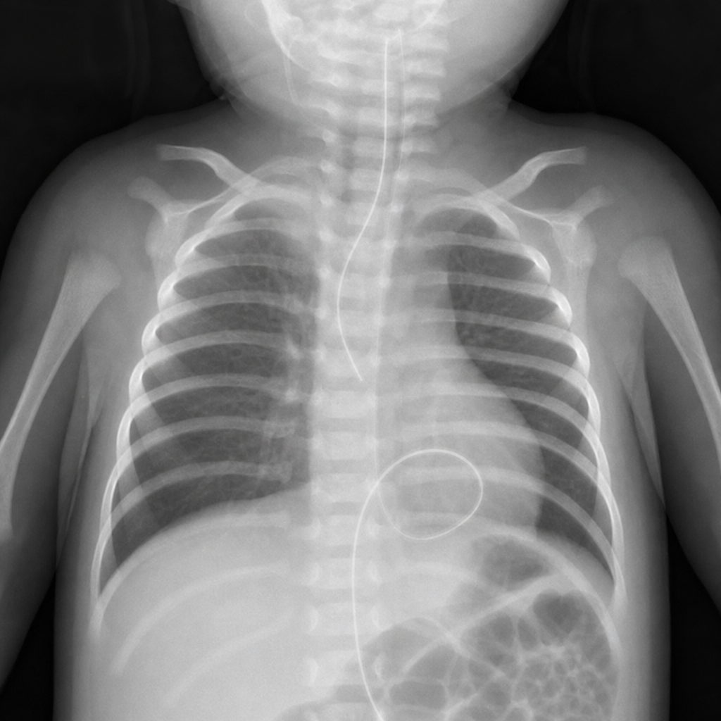
A term newborn presents with frothing, excessive drooling, and coughing with feeds. There was a history of polyhydramnios in the antenatal period. A chest X-ray was done showing a specific finding. What is the diagnosis?
Bag and mask ventilation is contraindicated in all except?
Which of the following is NOT true regarding the indications for exchange transfusion in neonates?
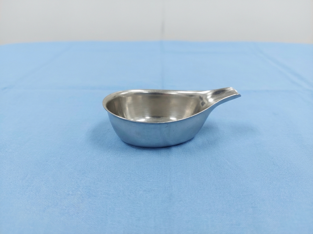
The item shown below is used to feed newborns born at what gestational age?
A 4-day-old infant, delivered at home, is brought to the emergency with complaints of bleeding from the umbilical stump. His PT is 24 seconds, APTT is 37 seconds, and platelet count is 200,000/mm3. Apart from maintaining intravascular volume, what would be the definitive line of management?
A three-day-old term infant, born at home and exclusively breast-fed, presented with lethargy, bulging fontanel, and bright red blood from the rectum. What is the most likely etiology of this condition?
Practice by Chapter
Neonatal Resuscitation
Practice Questions
Care of the Normal Newborn
Practice Questions
Prematurity and Low Birth Weight
Practice Questions
Respiratory Distress Syndrome
Practice Questions
Neonatal Jaundice
Practice Questions
Neonatal Sepsis
Practice Questions
Necrotizing Enterocolitis
Practice Questions
Intraventricular Hemorrhage
Practice Questions
Persistent Pulmonary Hypertension
Practice Questions
Perinatal Asphyxia
Practice Questions
Neonatal Seizures
Practice Questions
Congenital Anomalies
Practice Questions
Want unlimited practice?
Get full access to all questions, explanations, and performance tracking.
Scan to download app