
Neonatology — MCQs

On this page
Fetal alcohol syndrome is characterized by which of the following features?
Which of the following is NOT a risk factor for neonatal hyperbilirubinemia?
Which component is NOT included in the standard APGAR score, considering its historical 'A/E' (Appearance/Color) interpretation?
Children of parents with which of the following blood group combinations have the highest risk of erythroblastosis fetalis?
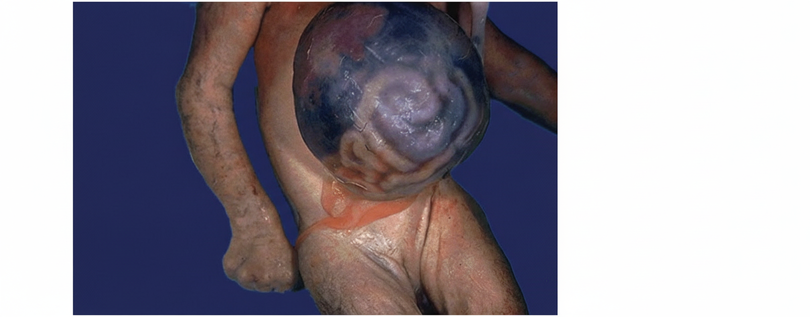
A neonate is found to have a condition as shown in the image. Which of the following is a common associated anomaly?
What is the APGAR score for a newborn with the following findings: Heart Rate 120/min, Respiratory Rate 40/min, strong respiratory efforts, peripheral cyanosis, grimace during suctioning, slightly flexed posture, and no active limb movements?
After 3 days of birth, the base of the umbilical cord is red and swollen. What does this indicate?
A newborn presents with jaundice within the first 24 hours of life. The mother's blood group is O positive. What is the next line of management?
Bronze baby syndrome is due to which of the following?
Antibodies for which of the following conditions are NOT transmitted from mother to fetus?
Practice by Chapter
Neonatal Resuscitation
Practice Questions
Care of the Normal Newborn
Practice Questions
Prematurity and Low Birth Weight
Practice Questions
Respiratory Distress Syndrome
Practice Questions
Neonatal Jaundice
Practice Questions
Neonatal Sepsis
Practice Questions
Necrotizing Enterocolitis
Practice Questions
Intraventricular Hemorrhage
Practice Questions
Persistent Pulmonary Hypertension
Practice Questions
Perinatal Asphyxia
Practice Questions
Neonatal Seizures
Practice Questions
Congenital Anomalies
Practice Questions
Want unlimited practice?
Get full access to all questions, explanations, and performance tracking.
Scan to download app