A 3-4 month old baby with heart rate 250/min, QRS complex less than 0.07 sec and no P wave, Diagnosis will be :
What is the appropriate management for a neonate presenting with vaginal bleeding?
Most common cause of interventricular bleed is?
Earliest clinical sign of raised intracranial pressure is:
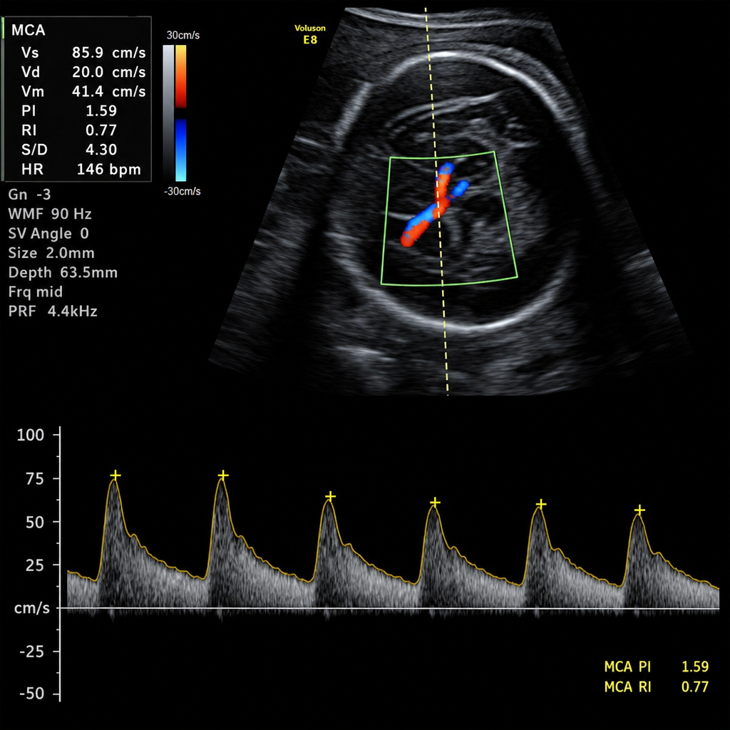
Which of the following medications is contraindicated in head trauma patients?
What is the preferred management for patent ductus arteriosus (PDA) in a preterm infant?
Which condition is associated with exclusively fetal blood loss?
A child with moderate to severe head injury is admitted in PICU. First line treatments are all except:
Following are the features of raised intracranial tension except -
A newborn presents with subconjunctival hemorrhage. The treatment is
Explanation: **SVT** - A heart rate of 250/min in a 3-4 month old infant, along with a **narrow QRS complex (<0.07 sec)**, is highly indicative of **supraventricular tachycardia (SVT)**. - The **absence of visible P waves** suggests that the atrial activity is either too rapid to be clearly distinguished or is retrograde and hidden within the QRS complex. *VT* - **Ventricular tachycardia (VT)** is characterized by **wide QRS complexes** (typically >0.09-0.10 sec in adults, proportionally less in infants) because the impulse originates in the ventricles. - The patient's QRS complex is **narrow (<0.07 sec)**, ruling out typical VT. *PSVT with block* - **Paroxysmal supraventricular tachycardia (PSVT) with block** would still present with a rapid atrial rate, and while there might be block to the ventricles, the dominant rhythm would stem from supraventricular activity causing narrow QRS, but the term "with block" usually implies some degree of AV nodal block which would lead to a ventricular rate slower than the atrial rate, unlike the observed 250/min. - The absence of P waves makes identification of a specific "block" pattern difficult, and the high ventricular rate favors a direct conduction rather than a blocked rhythm limiting ventricular response. *Sinus tachycardia* - **Sinus tachycardia** is usually characterized by discernible **P waves** preceding each QRS complex and a heart rate that typically doesn't exceed 220 bpm in infants unless under extreme physiological stress. - A heart rate of 250/min is generally above the physiological limit for sinus tachycardia in infants, and the **absence of P waves** further distinguishes it from sinus tachycardia.
Explanation: ***Observation and reassurance*** - **Neonatal vaginal bleeding** is often a benign condition caused by the withdrawal of maternal hormones (typically **estrogen**) after birth. - It usually resolves spontaneously within a few days or weeks, requiring no active medical intervention. *Administration of estrogen* - Administering estrogen would be inappropriate as the bleeding is caused by the withdrawal of maternal estrogen, not a deficiency. - Exogenous estrogen could further disrupt the delicate hormonal balance in the neonate. *Administration of progesterone* - Progesterone is largely irrelevant to the physiological mechanisms behind neonatal vaginal bleeding, which are primarily related to estrogen withdrawal. - There is no medical indication for progesterone treatment in this context. *Infusion of cryoprecipitate* - **Cryoprecipitate** contains clotting factors and is used to treat **bleeding disorders** or **DIC** where specific factor deficiencies are present. - There is no indication of a coagulation disorder in a neonate with physiological vaginal bleeding.
Explanation: ***Parenchymal bleed*** - A **parenchymal bleed**, particularly one that is large or occurs near the ventricles, can rupture into the ventricular system, leading to an **interventricular hemorrhage (IVH)** [2], [3]. - This is the most frequent cause of secondary IVH in adults, often due to conditions like **hypertensive hemorrhage** [3] or **arteriovenous malformations (AVMs)** within the brain parenchyma [2]. *SAH* - **Subarachnoid hemorrhage (SAH)** involves bleeding into the space between the arachnoid and pia mater, most commonly due to a ruptured aneurysm. - While SAH can sometimes track into the ventricles, it's not the primary or most common cause of significant interventricular bleeding compared to direct rupture from a parenchymal source [2]. *EDH* - **Epidural hemorrhage (EDH)** is typically caused by trauma, leading to bleeding between the dura mater and the skull [1]. - It is located entirely outside the brain parenchyma and the ventricular system, making it an unlikely direct cause of interventricular bleed. *SDH* - **Subdural hemorrhage (SDH)** involves bleeding between the dura and arachnoid mater, often due to tearing of bridging veins, also primarily from trauma [3]. - Like EDH, SDH is extra-axial and generally does not directly cause interventricular bleeding.
Explanation: ***Altered behavior*** - The earliest clinical sign of **raised intracranial pressure (ICP)** is often subtle changes in **mental status** and **behavior**, such as **restlessness**, irritability, or confusion. [1] - These changes reflect the brain's initial response to increasing pressure before more overt neurological signs develop. [1] *Hypotension* - **Hypotension** is generally not an early sign of raised ICP and can even be a late finding, particularly if a **Cushing reflex (hypertension, bradycardia, irregular breathing)** is developing. - Decreased blood pressure is more often associated with **shock** or other causes of systemic instability. *Tachycardia* - **Tachycardia** is not an early indicator of raised ICP; in fact, the **Cushing reflex**, a late sign of severe ICP elevation, is characterized by **bradycardia**. [1] - Tachycardia might indicate other issues like **hypovolemia** or **anxiety**. *Dyspnea* - **Dyspnea (difficulty breathing)** is a significant but typically **late sign** of raised ICP, often indicating brainstem compression and disruption of respiratory centers. [1] - It would usually be preceded by alterations in consciousness and other neurological deficits.
Explanation: ***Corticosteroids*** - While previously used, **corticosteroids** are now contraindicated in traumatic brain injury (TBI) due to evidence suggesting they may increase mortality. - **CRASH trial** showed that corticosteroids increased the risk of death in patients with head injury, possibly by exacerbating secondary brain injury. *Furosemide* - **Furosemide** can be used in certain situations to reduce intracranial pressure by inducing diuresis and reducing cerebral edema, especially when combined with mannitol. - It works by inhibiting the Na-K-2Cl cotransporter in the thick ascending limb of the loop of Henle, leading to increased water excretion. *Thiopentone* - **Thiopentone** (a barbiturate) can be used in severe head trauma to reduce cerebral metabolic rate, thereby decreasing cerebral blood flow and intracranial pressure. - It induces a **pharmacological coma** and provides neuroprotection by scavenging free radicals and stabilizing cell membranes. *Mannitol* - **Mannitol** is an osmotic diuretic commonly used to reduce intracranial pressure in head trauma by creating an osmotic gradient that draws water out of the brain parenchyma. - It is administered intravenously and works rapidly to decrease brain volume and improve cerebral perfusion pressure.
Explanation: ***IV Indomethacin*** - **Indomethacin** is a **prostaglandin synthesis inhibitor** that promotes the constriction and closure of the patent ductus arteriosus. - It is preferred due to its effectiveness in closing PDA non-invasively in preterm infants. *Surgical ligation* - This is an **invasive procedure** reserved for cases where medical management with indomethacin fails or is contraindicated. - While effective, it carries surgical risks such as **infection** and potential **vocal cord paralysis**. *Diuretics* - **Diuretics** are used to manage **pulmonary edema** or **heart failure symptoms** associated with a large PDA by reducing fluid overload. - They do not directly cause the closure of the patent ductus arteriosus itself. *Oxygen therapy* - **Oxygen therapy** is crucial for managing respiratory distress and maintaining adequate oxygen saturation in preterm infants. - However, oxygen can sometimes *inhibit* ductal closure in preterm infants by reducing pulmonary vascular resistance, and therefore, it is not the primary intervention for PDA closure.
Explanation: ***Vasa previa*** - Vasa previa occurs when **fetal blood vessels** from the umbilical cord traverse the membranes over the cervical os, underneath the fetal presenting part. - Rupture of these unprotected vessels, which can happen during labor or membrane rupture, leads to **exclusively fetal blood loss**, posing a high risk of fetal exsanguination and death. *Placenta praevia* - This condition involves the **placenta implanting low** in the uterus, potentially covering the internal cervical os. - Bleeding in placenta previa is typically **maternal** in origin, resulting from the detachment of the placenta from the uterine wall as the cervix dilates. *Polyhydramnios* - Polyhydramnios is characterized by an **excessive amount of amniotic fluid**. - It is not directly associated with antepartum or intrapartum bleeding, but rather with conditions that affect fetal swallowing or urination, such as **fetal gastrointestinal anomalies** or maternal diabetes. *Oligohydramnios* - Oligohydramnios refers to an **insufficient amount of amniotic fluid**. - While it can be associated with various fetal and maternal complications, such as **renal agenesis** or premature rupture of membranes, it does not typically cause blood loss.
Explanation: ***IV mannitol*** - While **intravenous mannitol** is used in the management of head injury to reduce **intracranial pressure (ICP)**, it is **not a first-line treatment**. - It is a **second-line therapy** reserved for documented or suspected elevated ICP despite initial supportive measures. - First-line management focuses on maintaining adequate oxygenation, ventilation, and cerebral perfusion, while mannitol is used for specific ICP management when needed. *Analgesia and sedation* - **Analgesia and sedation** are essential **first-line treatments** to reduce pain, anxiety, and agitation, which can increase **intracranial pressure (ICP)**. - These therapies ensure patient comfort, decrease metabolic demand, facilitate mechanical ventilation, and prevent secondary brain injury. *Hypothermia* - **Therapeutic hypothermia** is **NOT routinely recommended** as a first-line treatment in pediatric traumatic brain injury. - Current evidence (including the Cool Kids trial) has not demonstrated benefit, and it may be associated with adverse effects. - It is considered **investigational** and not part of standard first-line management protocols. - **Note**: While this is also not first-line, the question specifically tests knowledge that mannitol is second-line therapy for ICP management. *Controlled mechanical ventilation* - **Controlled mechanical ventilation** is a fundamental **first-line treatment** for severe head injury to secure the airway and ensure adequate oxygenation and ventilation. - Prevents secondary brain injury from **hypoxia** and **hypercapnia**, which can worsen outcomes. - Maintaining appropriate **PaCO2 levels** is critical to control cerebral blood flow and intracranial pressure.
Explanation: ***Tachycardia*** - **Tachycardia** (increased heart rate) is generally *not* a direct feature of raised intracranial tension (ICT); rather, the classic cardiovascular response is **bradycardia** (decreased heart rate) as part of the Cushing's reflex [1]. - The Cushing's reflex, triggered by increased ICP, involves hypertension, bradycardia, and irregular respirations, a protective mechanism to maintain cerebral perfusion. *Convulsions* - **Convulsions** can occur due to focal brain irritation or global cerebral dysfunction caused by severe or rapidly rising intracranial pressure. - Elevated ICP can compromise neuronal function and integrity, leading to abnormal electrical activity. *Papilloedema* - **Papilloedema** (swelling of the optic disc) is a classic and frequently observed sign of chronic or sustained raised intracranial tension [2]. - It results from the obstruction of axoplasmic flow and venous return in the optic nerve due to increased pressure in the subarachnoid space surrounding the optic nerve. *Altered sensorium* - **Altered sensorium**, ranging from confusion and disorientation to stupor and coma, is a common and serious manifestation of raised intracranial tension [1]. - It occurs due to diffuse cerebral dysfunction as the brain is compressed and its blood supply is compromised.
Explanation: ***No treatment*** - **Subconjunctival hemorrhage** in a newborn is typically **benign** and **resolves spontaneously** within **1-2 weeks**. - It is often caused by the trauma of birth and does not require intervention. *Antibiotic eye drops* - These are indicated for **bacterial conjunctivitis** or to prevent bacterial infection, which is not the case here. - Using antibiotics without a bacterial indication is unnecessary and can contribute to **antibiotic resistance**. *Aspiration* - **Aspiration** is an invasive procedure and is **not indicated** for a subconjunctival hemorrhage, which is a collection of blood under the conjunctiva. - It could cause further damage or introduce infection. *Antibiotic and steroid drops* - **Steroid drops** are typically used to reduce **inflammation**, which is not the primary issue in a subconjunctival hemorrhage. - Like plain antibiotic drops, the **antibiotic component** is not necessary in the absence of infection.
Get full access to all questions, explanations, and performance tracking.
Start For Free