
Infectious Diseases — MCQs

On this page
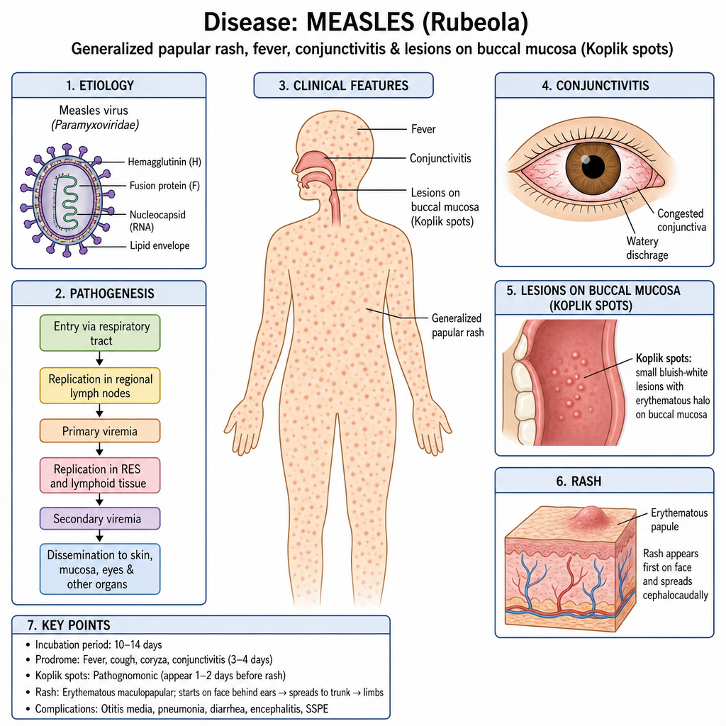
In which disease a generalized papular rash, fever, conjunctivitis & lesion on buccal mucosa shown below are seen?
Complications of measles are all except:
Koplik spots are characteristic of:-
Reye's syndrome following influenza is most commonly associated with -
In a small child diagnosed with H. influenzae meningitis, what investigation must be done before discharging him from the hospital?
Which of the following is NOT a feature of HIV infection in childhood -
A 10-year-old girl presented with fever, convulsions, and neck rigidity. CSF findings are protein 150 mg/dL, sugar 40 mg/dL with lymphocytic pleocytosis –
Pathognomonic of measles?
A child who was treated for H. influenzae meningitis is being discharged. Most important investigation to be done before discharge is:
Most serious characteristic finding of HIV in children is?
Practice by Chapter
Vaccine-Preventable Diseases
Practice Questions
Immunization Schedule
Practice Questions
Common Childhood Infections
Practice Questions
Pediatric HIV
Practice Questions
Congenital Infections
Practice Questions
Fever in Infants and Children
Practice Questions
Meningitis and Encephalitis
Practice Questions
Respiratory Tract Infections
Practice Questions
Gastrointestinal Infections
Practice Questions
Parasitic Infections
Practice Questions
Tuberculosis in Children
Practice Questions
Opportunistic Infections
Practice Questions
Want unlimited practice?
Get full access to all questions, explanations, and performance tracking.
Scan to download app