
Infectious Diseases — MCQs

On this page
Which one of the following is the recommended site for immunization with hepatitis B vaccine in young children for ensuring reliable absorption?
Which statement about congenital syphilis is FALSE?
A child presents with complaints of fever, rash, body ache, and throat ache. He had a history of thorn prick injury a week back. What antibiotics would you give empirically to this child?
A 10 week old child comes for vaccination, with previous history of inconsolable cry & fever after getting vaccinated at 6 weeks. What should be done next?
A 10-week-old baby came for vaccination. The baby had a previous history of inconsolable crying and fever (40°C) after vaccination at 6 weeks. What should be given now?
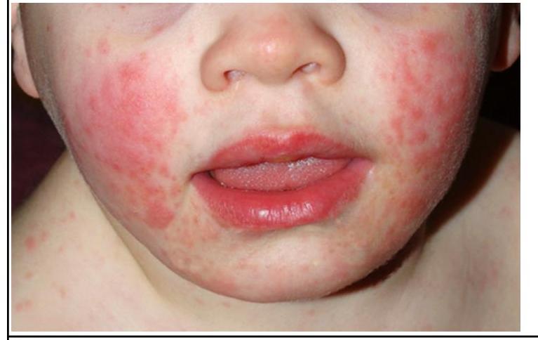
A child presenting with the following appearance is at risk of developing?
A child presents with fever and vesicular lesions on the upper limb and the lower limb. Neck stiffness was present. Similar lesions were present on the palms, soles, and oral cavity. CSF analysis revealed normal glucose levels and elevated lymphocytes and protein. What is the most likely diagnosis?
Which of the following is given as a part of the therapy for a child with severe COVID?
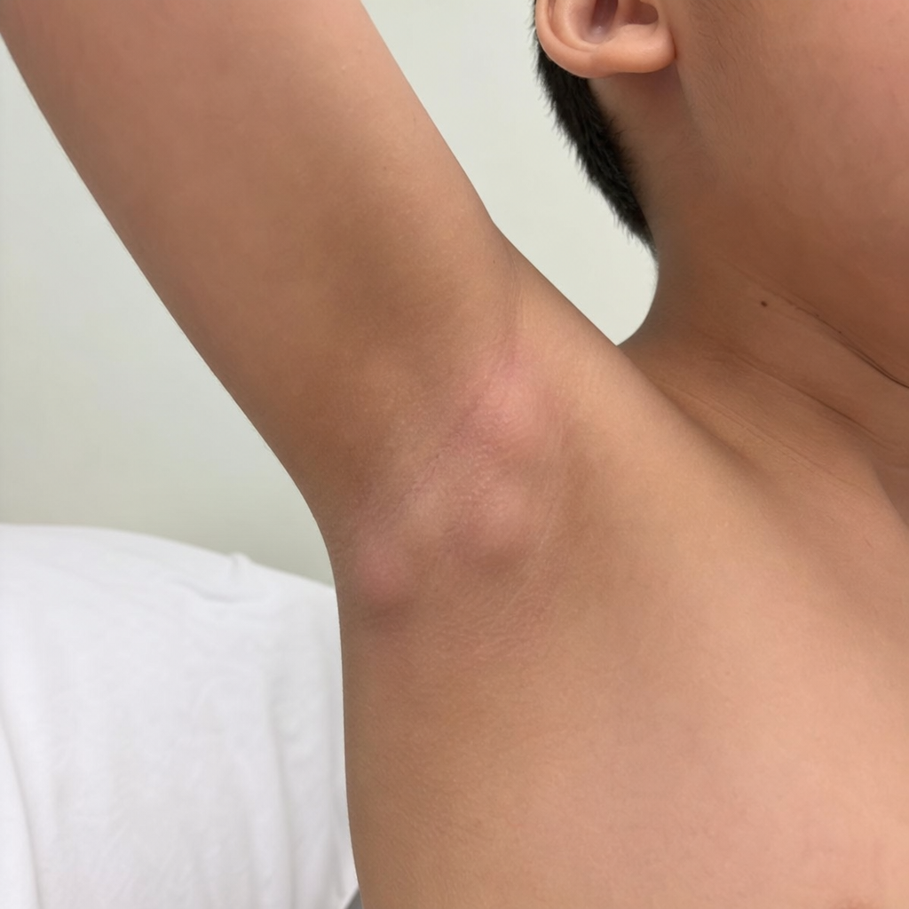
A 10-year-old boy is brought to the physician with painful and enlarged lymph nodes in his right axilla that was noticed 5 days ago and has slowly grown bigger. He has had weakness, sweating, and poor appetite during this time. The boy was born at 39 weeks gestation via spontaneous vaginal delivery. He is up to date on all vaccines and is meeting all developmental milestones. He does not take any medication. There are no similar cases in the family. On physical exam, his temperature is 38.2°C (100.8°F), the pulse is 89/min, the respiratory rate is 13/min, and the blood pressure is 110/60 mm Hg. In his right axilla, there are multiple tender, fluctuant, and enlarged lymph nodes with overlying erythematous skin. There is a separate lesion on the child's forearm (see image). The lesion is painless to palpation and appears inflamed. Additional history should be obtained regarding which of the following?
For a child 3-5 months of age with H1N1, treatment Oseltamivir dose is
Practice by Chapter
Vaccine-Preventable Diseases
Practice Questions
Immunization Schedule
Practice Questions
Common Childhood Infections
Practice Questions
Pediatric HIV
Practice Questions
Congenital Infections
Practice Questions
Fever in Infants and Children
Practice Questions
Meningitis and Encephalitis
Practice Questions
Respiratory Tract Infections
Practice Questions
Gastrointestinal Infections
Practice Questions
Parasitic Infections
Practice Questions
Tuberculosis in Children
Practice Questions
Opportunistic Infections
Practice Questions
Want unlimited practice?
Get full access to all questions, explanations, and performance tracking.
Scan to download app