AIDS, secondary infection will be all except
CD40 deficiency in a person signifies?
Which of the following methods is NOT recommended for the diagnosis of HIV infection in a 2-month-old child?
With the lack of CD40 in B cells, which immunological abnormality is seen?
Which of the following is not a complication of Congenital Rubella Syndrome (CRS)?
According to WHO, the major clinical sign of HIV infection in children in stage 1 is the following.
Given the immunologic abnormalities of normal serum IgG, normal serum IgA, normal serum IgM, decreased T-cell function, and decreased parathyroid function, which clinical presentation is most likely?
Best method to diagnose HIV in an infant?
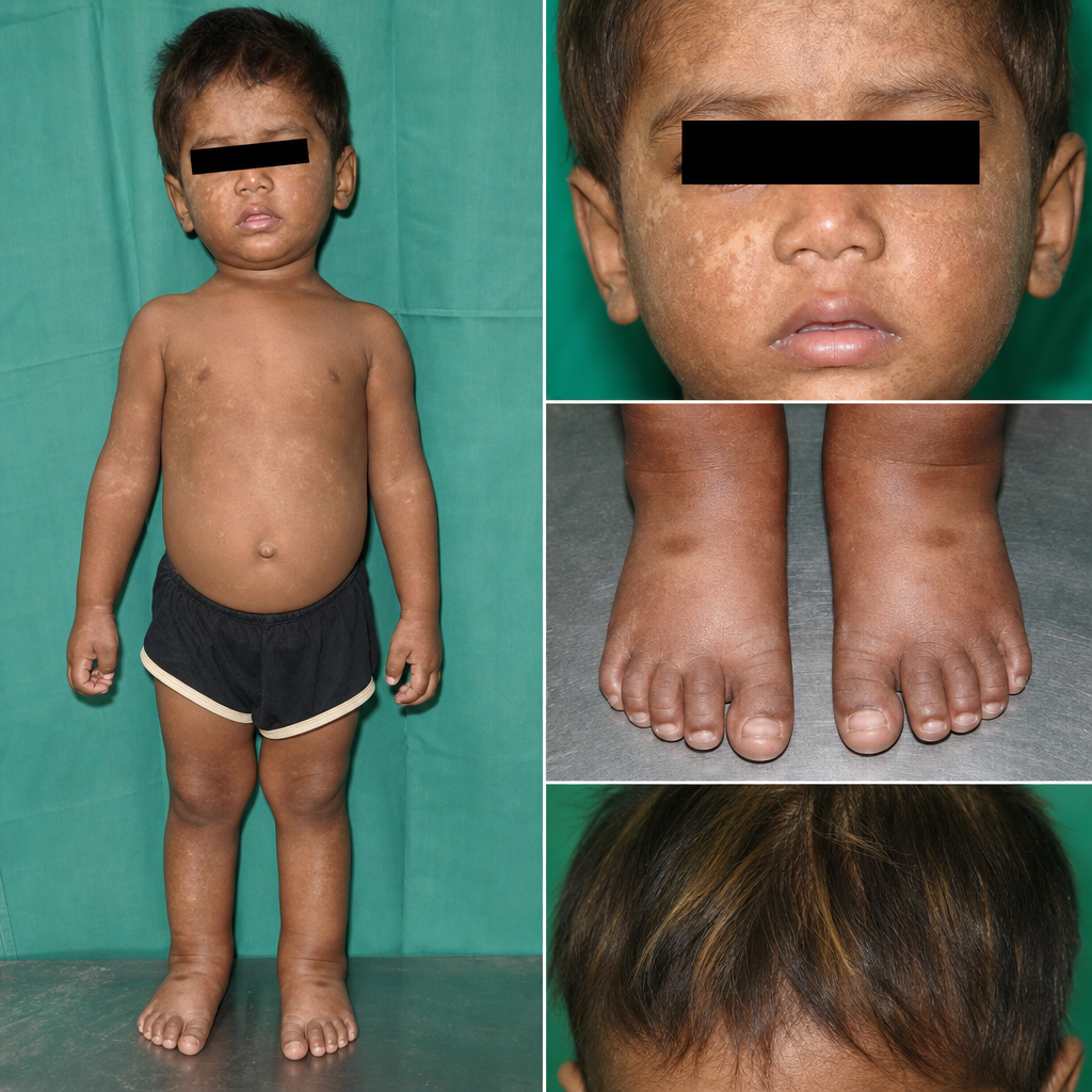
What is a characteristic feature of Systemic Juvenile Idiopathic Arthritis?
A patient presents with thrombocytopenia, eczema, and recurrent infections. What is the most probable diagnosis?
Explanation: ***Kaposi's sarcoma*** - Kaposi's sarcoma is a **cancer** caused by human herpesvirus 8 (HHV-8) [2] that is common in patients with AIDS, but it is a **malignancy**, not a secondary infection [2],[3]. - While it arises due to immune suppression, it represents abnormal cell proliferation rather than direct microbial invasion. *Candida* - **Candidiasis** (e.g., oral thrush, esophageal candidiasis) is a common opportunistic fungal infection in AIDS patients due to their **impaired cellular immunity** [1]. - It often presents as **white plaques** on mucous membranes and is a clear example of a secondary infection. *HSV* - **Herpes Simplex Virus (HSV)** infections, including oral and genital herpes, are common and often severe in AIDS patients. - Due to immunocompromise, these infections can be **more widespread**, chronic, or recur frequently, qualifying as secondary infections. *Rubella* - **Rubella (German measles)** is a viral infection that is generally mild and self-limiting in immunocompetent individuals. - It is **not considered an opportunistic infection** or a common secondary infection specifically associated with AIDS; rather, it is listed as a differential diagnosis for the primary HIV infection rash [1].
Explanation: ***IgM increase*** - A deficiency in **CD40**, or its ligand **CD40L** (found on T helper cells), disrupts **T-cell-dependent B cell activation** and **class switching**. - Without proper signaling through CD40/CD40L, B cells cannot undergo **isotype switching** from **IgM** to IgG, IgA, or IgE, leading to elevated IgM levels and deficiencies in other antibody classes. *IgG increase* - **IgG levels** would likely be **decreased** in CD40 deficiency due to the impaired ability of B cells to undergo **class switching** from IgM to other antibody isotypes. - The primary role of CD40/CD40L interaction is to facilitate this class switching process. *T cell absent* - **CD40 deficiency** does not directly cause the absence of **T cells**; rather, it affects the ability of T cells to adequately activate B cells. - T-cell absence or severe dysfunction would be indicative of a different primary immunodeficiency, such as **SCID (Severe Combined Immunodeficiency)**. *B cell absent* - **CD40 deficiency** does not result in the absence of **B cells**; B cells are present but are dysfunctional in terms of antibody class switching. - Conditions like **X-linked agammaglobulinemia (XLA)** are characterized by the absence or severe deficiency of B cells.
Explanation: ***HIV ELISA*** - **HIV ELISA** (Enzyme-linked Immunosorbent Assay) detects **HIV antibodies**, which are maternally derived and can persist in newborns for up to 18 months, leading to **false positive** results. - Therefore, antibody-based tests are **not suitable** for diagnosing HIV infection in infants under 18 months of age. *Viral culture* - **HIV viral culture** can directly detect the presence of replication-competent virus in an infant's blood. - While sensitive, it is **expensive**, labor-intensive, and takes a long time (several weeks) to obtain results, making it less practical for routine diagnosis. *DNA-PCR* - **DNA PCR (Polymerase Chain Reaction)** directly detects **HIV proviral DNA** within infected cells, making it highly specific and sensitive for early infant diagnosis. - It is currently the **recommended method** for HIV diagnosis in infants and young children, especially in the first few months of life. *P24 antigen assay* - The **P24 antigen assay** detects the **core protein of the HIV virus**, indicating active viral replication. - It can be used for early diagnosis in infants but may be less sensitive than DNA PCR, particularly in the presence of maternal antibodies or during early infection.
Explanation: ***Decreased IgG and increase in IgM*** - The interaction between **CD40 on B cells** and **CD40L (CD154) on T helper cells** is crucial for **B cell activation**, proliferation, and **class switch recombination** (CSR). - Without this interaction, B cells cannot undergo CSR, leading to a failure to produce **IgG, IgA, or IgE**, while **IgM levels remain high** because IgM production is the initial default. *Total lack of NK cells* - **Natural Killer (NK) cells** are part of the innate immune system and their development is largely independent of CD40-CD40L signaling. - The absence of CD40 on B cells primarily affects adaptive humoral immunity, not NK cell numbers or function. *Lack of CD8 mediated cytotoxicity* - **CD8+ T cells** mediate cytotoxicity against infected or cancerous cells and their activation is primarily dependent on antigen presentation by **MHC class I molecules** and costimulation, not directly on B cell CD40. - While B cells can act as APCs, their CD40 interaction is more critical for T helper cell help for humoral responses. *Inability of neutrophil against infections* - **Neutrophils** are phagocytic cells important in innate immunity, and their function is largely independent of CD40 on B cells. - Neutrophil activity relies on pathogen recognition, phagocytosis, and degranulation, which are not directly regulated by the B cell CD40-CD40L pathway.
Explanation: ***Macrocephaly*** - While CRS can lead to various neurological complications, **macrocephaly** (abnormally large head circumference) is not a typical manifestation of the syndrome. Neurological issues in CRS more commonly involve **microcephaly** due to brain damage. - Other common neurological complications include **meningoencephalitis** and developmental delays, but not an enlarged head. *Retinopathy* - **Pigmentary retinopathy** (salt-and-pepper retinopathy) is a classic ocular manifestation of CRS, often present at birth. - This is a direct consequence of the rubella virus affecting the developing retinal structures. *Spontaneous abortion* - Maternal rubella infection, especially during the **first trimester**, carries a significant risk of **spontaneous abortion** due to severe fetal damage. - The virus's teratogenic effects can be so profound that the fetus is not viable. *Cardiac abnormalities* - **Congenital heart defects** are a hallmark of CRS, with **patent ductus arteriosus (PDA)** and **pulmonary artery stenosis** being the most common. - These abnormalities result from the rubella virus interfering with normal cardiac development during embryogenesis.
Explanation: ***Generalised lymphadenopathy*** - According to WHO staging for HIV infection in children, **persistent generalized lymphadenopathy (PGL)** is a major clinical sign in Stage 1. - This stage is characterized by asymptomatic or mild clinical manifestations, with PGL being one of the key visible indicators. - PGL is defined as enlarged lymph nodes (>1 cm) in two or more non-contiguous sites, excluding inguinal nodes. *Not gaining weight* - **Failure to thrive** or **unexplained moderate malnutrition** typically falls under WHO clinical Stage 2 or 3, not Stage 1. - While weight loss can occur in HIV, it usually signifies more advanced disease progression in children. *Oral candidiasis* - **Oral candidiasis** in children with HIV usually indicates more advanced disease and is classified under **WHO clinical Stage 2 or 3**, depending on its persistence and severity. - It suggests a compromised immune system beyond the earliest stages of HIV infection. *None of the options* - This option is incorrect because **generalized lymphadenopathy** is indeed a major clinical sign of HIV infection in children in Stage 1, according to WHO criteria. - The other options provided represent signs found in later stages of HIV disease in children.
Explanation: ***A distinctive-appearing 8-month-old boy with an interrupted aortic arch, hypocalcemia, and cleft palate*** - This presentation is highly suggestive of **DiGeorge syndrome**, characterized by **thymic hypoplasia** (leading to decreased T-cell function) and **parathyroid hypoplasia** (causing hypocalcemia). - **Cardiac defects** (like an interrupted aortic arch) and **facial anomalies** (including cleft palate) are also classic features of this disorder, which involves a deletion on chromosome 22q11.2. *A 1-year-old boy with severe eczema, recurrent middle-ear infections, lymphopenia, and thrombocytopenia* - This clinical picture describes **Wiskott-Aldrich syndrome**, an X-linked disorder characterized by the triad of eczema, thrombocytopenia (with small platelets), and immunodeficiency leading to recurrent infections. - While it involves immunodeficiency and lymphopenia, it does not typically present with decreased parathyroid function. *A 9-year-old boy with an eczema-like rash and recurrent severe staphylococcal infections* - This presentation is characteristic of **hyper-IgE syndrome** (Job's syndrome), an immunodeficiency characterized by extremely elevated IgE levels, recurrent staphylococcal skin infections, and eczema. - The immunologic abnormalities described in the stem (normal Ig levels, decreased T-cell function, decreased parathyroid function) do not match the key features of hyper-IgE syndrome. *A 5-year-old boy who, after 3 months of age, developed recurrent otitis media, pneumonia, diarrhea, and sinusitis, often with simultaneous infections at two or more disparate sites* - This description is consistent with **X-linked agammaglobulinemia (XLA)**, where B-cell maturation is blocked, leading to a profound deficiency of all immunoglobulin classes. - The stem mentions normal serum IgG, IgA, and IgM, which rules out XLA.
Explanation: ***PCR*** - **Polymerase Chain Reaction (PCR)** detects **HIV nucleic acids** (DNA or RNA) directly, which is crucial for infants because maternal antibodies can persist for up to 18 months, interfering with antibody-based tests. - PCR allows for early diagnosis, often within the first few weeks or months of life, facilitating timely intervention. *ELISA* - **Enzyme-linked immunosorbent assay (ELISA)** detects HIV antibodies. - In infants, ELISA can be misleading due to the presence of **maternal HIV antibodies** transferred across the placenta, making it unreliable for diagnosing active infection. *Western blot* - **Western blot** is used to confirm positive ELISA results in adults by detecting specific HIV proteins. - Like ELISA, it relies on the detection of **antibodies** and is therefore not reliable in infants due to maternally transmitted antibodies. *All of the options* - This option is incorrect because **ELISA** and **Western blot** are antibody-based tests that are unreliable in infants due to the presence of **maternal antibodies**. - Only **PCR** directly detects the virus itself, making it the preferred diagnostic method in this age group.
Explanation: ### Explanation **Systemic Juvenile Idiopathic Arthritis (sJIA)**, also known as Still’s disease, is a unique subtype of JIA characterized by prominent extra-articular features. **Why the correct answer is right:** In sJIA, the **Rheumatoid Factor (RF) is characteristically negative**. Unlike the polyarticular subtype (which can be RF positive), sJIA is considered an autoinflammatory disease rather than a classic autoimmune disease. Diagnosis is clinical, based on the presence of arthritis in one or more joints associated with (or preceded by) a fever of at least 2 weeks' duration that is daily ("quotidian") for at least 3 days, accompanied by features like an evanescent salmon-pink rash, lymphadenopathy, or serositis. **Analysis of Incorrect Options:** * **A. Uveitis is a feature:** This is incorrect for sJIA. Chronic anterior uveitis is a classic complication of **Oligoarticular JIA** (especially if ANA positive). Uveitis is very rare in the systemic subtype. * **B. It occurs after 16 years of age:** By definition, JIA must have an onset **before the age of 16**. If similar symptoms occur after 16, it is termed Adult-Onset Still’s Disease (AOSD). * **C. NSAIDs are contraindicated:** This is false. NSAIDs are often the **first-line** symptomatic treatment for pain and fever in JIA, though systemic steroids or biologics (IL-1 and IL-6 inhibitors) are usually required for definitive control. **High-Yield Clinical Pearls for NEET-PG:** * **Fever Pattern:** Classic "Quotidian" fever (spikes once daily, usually in the evening, returning to baseline). * **Laboratory Markers:** Marked leukocytosis, thrombocytosis, and highly elevated ESR/CRP. * **Ferritin:** Extremely high ferritin levels are common and can signal the onset of **Macrophage Activation Syndrome (MAS)**, a life-threatening complication of sJIA. * **Biologics of Choice:** Tocilizumab (IL-6 inhibitor) and Anakinra/Canakinumab (IL-1 inhibitors).
Explanation: **Explanation:** The classic triad of **thrombocytopenia, eczema, and recurrent infections** is the hallmark presentation of **Wiskott-Aldrich Syndrome (WAS)**. 1. **Why A is Correct:** WAS is an X-linked recessive disorder caused by a mutation in the *WASp* gene, which leads to defects in the actin cytoskeleton of hematopoietic cells. This results in: * **Thrombocytopenia:** Characteristically presents with **micro-platelets** (small size), leading to bleeding tendencies (e.g., petechiae, melena). * **Eczema:** Typically develops within the first year of life. * **Immunodeficiency:** Defects in both T-cells and B-cells lead to recurrent infections with encapsulated bacteria and opportunistic pathogens. 2. **Why the others are Incorrect:** * **B. Agammaglobulinemia (Bruton’s):** Presents with recurrent pyogenic infections due to B-cell deficiency, but lacks thrombocytopenia and eczema. * **C. Chediak-Higashi Syndrome:** Characterized by **oculocutaneous albinism**, giant cytoplasmic granules in neutrophils, and peripheral neuropathy. * **D. Lazy Leukocyte Syndrome:** A defect in neutrophil chemotaxis and mobility; patients have neutropenia but not the classic triad of WAS. **High-Yield Clinical Pearls for NEET-PG:** * **Inheritance:** X-linked Recessive (mostly males). * **Lab Finding:** Low IgM, normal/high IgA and IgE, and **small-sized platelets** (pathognomonic). * **Complications:** High risk of **autoimmune hemolytic anemia** and **B-cell lymphomas**. * **Treatment:** Hematopoietic stem cell transplant (HSCT) is the definitive cure.
Get full access to all questions, explanations, and performance tracking.
Start For Free