A man had Egg omelet 1-4 hours back and developed diarrhea, vomiting and abdominal cramps. Most likely cause of poisoning is
Which type of dermatitis is evaluated through patch testing?
A man takes peanut and develops tongue swelling, neck swelling, stridor, hoarseness of voice. What is the probable diagnosis?
In chronic allergy, which Ig is more persistent in the body?
A 45-year-old patient with a known allergy to penicillin presents with an enterococcal endocarditis. The physician needs to prescribe an antibiotic but wants to ensure it is safe for a penicillin allergy. The patient has had previous allergic reactions to penicillin including rash & swelling. Which of the following drugs can be used safely in a patient allergic to penicillin?
Drug of choice for Enterococcus infection in a patient allergic to penicillin?
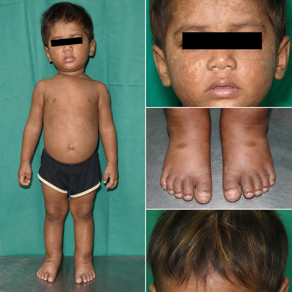
What is a characteristic feature of Systemic Juvenile Idiopathic Arthritis?
A patient presents with thrombocytopenia, eczema, and recurrent infections. What is the most probable diagnosis?
A child presents with delayed separation of the umbilical cord, leukocytosis, Down syndrome, and recurrent infections. What is the most likely diagnosis?
Which of the following is a feature of Henoch-Schönlein purpura?
Explanation: ***Staphylococcus*** - **Staphylococcal food poisoning** is characterized by a rapid onset (1-6 hours, most commonly 2-4 hours) of symptoms like **nausea**, **vomiting**, **abdominal cramps**, and **diarrhea** after consuming contaminated food. - Omelets prepared with eggs can be a source if handled improperly, allowing pre-formed **heat-stable enterotoxins** produced by *Staphylococcus aureus* to cause illness, even if the bacteria themselves are killed by cooking. *Bacillus cereus* - This organism can cause two types of food poisoning: diarrheal and emetic. The **emetic type**, characterized by prominent vomiting, has a rapid onset (0.5-6 hours, usually 1-5 hours) often associated with *rice products* contaminated with **heat-stable toxin**. - While vomiting is present, the consumption of an omelet and the typical 1-4 hour timeframe match Staphylococcal more closely, and *Bacillus cereus* diarrheal type usually has a longer incubation (6-15 hours). *Clostridium* - **Clostridium perfringens** food poisoning typically causes primarily **diarrhea** and **abdominal cramps** with less prominent vomiting, and has a longer incubation period of 6-24 hours. - **Clostridium botulinum** causes a neuroparalytic illness, not primarily gastroenteritis, and is associated with improperly canned foods. *Salmonella* - **Salmonella food poisoning** usually has a longer incubation period, typically 12-72 hours (average 12-36 hours), with symptoms including diarrhea, fever, and abdominal cramps. - The **rapid onset** (1-4 hours) in this case makes Salmonella less likely, as it primarily causes infection rather than pre-formed toxin ingestion.
Explanation: ***Contact dermatitis due to allergens*** - **Patch testing** is specifically used to identify specific **allergens** that trigger an **allergic contact dermatitis** reaction. - It involves applying suspected allergens to the skin and observing for a localized inflammatory response, indicating delayed type IV hypersensitivity. *Atopic dermatitis* - This is a chronic inflammatory skin condition characterized by **eczematous lesions** and severe **pruritus**, often linked to a genetic predisposition and immune dysfunction. - While allergy testing (e.g., prick tests, blood tests for IgE) might be used to identify triggers, **patch testing** is not the primary diagnostic tool for atopic dermatitis itself. *Irritant contact dermatitis* - This type of dermatitis is caused by direct **damage to the skin barrier** from exposure to caustic substances or irritants, not an immune-mediated allergic reaction. - Diagnosis is usually based on clinical history of exposure and symptom presentation, and **patch testing** is typically negative in these cases. *Discoid eczema* - Also known as **nummular dermatitis**, this condition presents with distinctive **coin-shaped lesions** and is often associated with dry skin or skin trauma. - Its etiology is generally unknown and not attributable to specific allergens detectable by **patch testing**.
Explanation: Andioneurotic edema - The combination of **tongue swelling**, **neck swelling**, **stridor**, and **hoarseness of voice** following peanut ingestion is highly suggestive of **angioneurotic edema**, a severe allergic reaction that can lead to airway obstruction [1]. - This is a life-threatening condition requiring immediate medical intervention, often associated with generalized **anaphylaxis** when triggered by allergens [2]. *FB in larynx* - While a **foreign body (FB) in the larynx** can cause stridor and hoarseness, the widespread swelling of the tongue and neck points away from a localized laryngeal obstruction [3]. - A laryngeal FB would typically be associated with a more sudden onset of choking and coughing, not diffuse edema [3]. *Parapharyngeal abscess* - A **parapharyngeal abscess** would typically present with **fever**, **severe throat pain**, and **trismus** (difficulty opening the mouth), which are not mentioned in this scenario. - The acute, rapid onset of symptoms after peanut consumption is inconsistent with the slower progression of an abscess. *FB bronchus* - A **foreign body in the bronchus** would primarily cause **coughing**, **wheezing**, and possibly **respiratory distress**, often unilateral, rather than severe global swelling of the tongue and neck. - Inspiratory stridor and hoarseness are more indicative of upper airway involvement than bronchial obstruction.
Explanation: ***Ig E*** - **IgE** is the primary antibody involved in **allergic reactions**, binding to receptors on **mast cells** and **basophils** to trigger histamine release. - In chronic allergy, sustained exposure to allergens leads to continuous production of IgE, making it a **persistent** and dominant immunoglobulin in the allergic response. *Ig A* - **IgA** is mainly found in **mucosal secretions**, such as tears, saliva, and gut, protecting against pathogens at these sites. - While important for immunity, IgA does not play a direct role in the **immediate hypersensitivity reactions** characteristic of chronic allergies. *Ig G* - **IgG** is the most abundant antibody in serum, providing **long-term immunity** against pathogens through neutralization, opsonization, and complement activation. - Though present, IgG is not the **primary mediator** of the **allergic response** in chronic allergy, instead often associated with protective immunity or certain non-IgE mediated hypersensitivities. *Ig M* - **IgM** is the first antibody produced during a **primary immune response** and is effective at activating the complement system. - It is predominantly found in the bloodstream and functions as a **short-term defender**, but it is not directly involved in the pathogenesis or persistence of chronic allergies.
Explanation: ***Vancomycin***- **Vancomycin** is a glycopeptide antibiotic that is **structurally unrelated to penicillin**, with no cross-reactivity in penicillin-allergic patients [2].- It has **excellent activity against Enterococcus species** and is the **preferred alternative for enterococcal endocarditis** in patients with penicillin allergy [1, 2].- Vancomycin provides reliable bactericidal activity against enterococci and is guideline-recommended for this indication in penicillin-allergic patients [1].*Aztreonam*- **Aztreonam** is a monobactam antibiotic with minimal cross-reactivity to penicillin allergies due to its unique beta-lactam structure.- However, aztreonam has **NO activity against Gram-positive organisms**, including Enterococcus species.- It would be **completely ineffective** for treating enterococcal endocarditis despite being safe in penicillin allergy.*Ceftriaxone*- **Ceftriaxone** is a third-generation cephalosporin that shares the beta-lactam ring structure with penicillins.- There is approximately **1-3% cross-reactivity risk** in patients with non-severe penicillin allergy, and up to 10% in those with severe reactions [1].- Given this patient's history of rash and swelling, ceftriaxone carries **unacceptable cross-reactivity risk** [1].*Piperacillin*- **Piperacillin** is an extended-spectrum penicillin antibiotic, belonging to the same drug class as penicillin [1].- It is **absolutely contraindicated** in penicillin-allergic patients due to identical allergenic epitopes [1].- Administration would carry a **high risk of severe allergic reaction**, including potential anaphylaxis [1].
Explanation: ***Vancomycin*** - **Vancomycin** is a glycopeptide antibiotic that is effective against **Gram-positive bacteria**, including *Enterococcus*, especially in patients with a **penicillin allergy**. - It inhibits **cell wall synthesis** by binding to the D-Ala-D-Ala terminus of peptidoglycan precursors, a different mechanism from penicillins. *Streptomycin* - **Streptomycin** is an aminoglycoside that inhibits **protein synthesis** and is primarily used in **combination therapy** for serious *Enterococcal* infections, but typically alongside a cell-wall active agent (like penicillin or vancomycin) for synergistic killing in endocarditis or other severe infections. - It is not usually recommended as a **monotherapy** for *Enterococcus*, especially in the context of penicillin allergy, as it doesn't provide bactericidal activity on its own against all enterococcal strains. *Cephalosporin* - **Cephalosporins** are **not active** against *Enterococcus spp.* as these bacteria intrinsically lack the **penicillin-binding proteins (PBPs)** that cephalosporins target effectively. - This **intrinsic resistance** makes cephalosporins an inappropriate choice for treating *Enterococcal* infections, regardless of penicillin allergy status. *Rifampicin* - **Rifampicin** is an antibiotic primarily used for **Mycobacterial infections** (e.g., tuberculosis) and some **Staphylococcal infections**, often in combination to prevent resistance. - It has **poor activity** against *Enterococcus* and is not a recommended treatment for *Enterococcal* infections.
Explanation: ### Explanation **Systemic Juvenile Idiopathic Arthritis (sJIA)**, also known as Still’s disease, is a unique subtype of JIA characterized by prominent extra-articular features. **Why the correct answer is right:** In sJIA, the **Rheumatoid Factor (RF) is characteristically negative**. Unlike the polyarticular subtype (which can be RF positive), sJIA is considered an autoinflammatory disease rather than a classic autoimmune disease. Diagnosis is clinical, based on the presence of arthritis in one or more joints associated with (or preceded by) a fever of at least 2 weeks' duration that is daily ("quotidian") for at least 3 days, accompanied by features like an evanescent salmon-pink rash, lymphadenopathy, or serositis. **Analysis of Incorrect Options:** * **A. Uveitis is a feature:** This is incorrect for sJIA. Chronic anterior uveitis is a classic complication of **Oligoarticular JIA** (especially if ANA positive). Uveitis is very rare in the systemic subtype. * **B. It occurs after 16 years of age:** By definition, JIA must have an onset **before the age of 16**. If similar symptoms occur after 16, it is termed Adult-Onset Still’s Disease (AOSD). * **C. NSAIDs are contraindicated:** This is false. NSAIDs are often the **first-line** symptomatic treatment for pain and fever in JIA, though systemic steroids or biologics (IL-1 and IL-6 inhibitors) are usually required for definitive control. **High-Yield Clinical Pearls for NEET-PG:** * **Fever Pattern:** Classic "Quotidian" fever (spikes once daily, usually in the evening, returning to baseline). * **Laboratory Markers:** Marked leukocytosis, thrombocytosis, and highly elevated ESR/CRP. * **Ferritin:** Extremely high ferritin levels are common and can signal the onset of **Macrophage Activation Syndrome (MAS)**, a life-threatening complication of sJIA. * **Biologics of Choice:** Tocilizumab (IL-6 inhibitor) and Anakinra/Canakinumab (IL-1 inhibitors).
Explanation: **Explanation:** The classic triad of **thrombocytopenia, eczema, and recurrent infections** is the hallmark presentation of **Wiskott-Aldrich Syndrome (WAS)**. 1. **Why A is Correct:** WAS is an X-linked recessive disorder caused by a mutation in the *WASp* gene, which leads to defects in the actin cytoskeleton of hematopoietic cells. This results in: * **Thrombocytopenia:** Characteristically presents with **micro-platelets** (small size), leading to bleeding tendencies (e.g., petechiae, melena). * **Eczema:** Typically develops within the first year of life. * **Immunodeficiency:** Defects in both T-cells and B-cells lead to recurrent infections with encapsulated bacteria and opportunistic pathogens. 2. **Why the others are Incorrect:** * **B. Agammaglobulinemia (Bruton’s):** Presents with recurrent pyogenic infections due to B-cell deficiency, but lacks thrombocytopenia and eczema. * **C. Chediak-Higashi Syndrome:** Characterized by **oculocutaneous albinism**, giant cytoplasmic granules in neutrophils, and peripheral neuropathy. * **D. Lazy Leukocyte Syndrome:** A defect in neutrophil chemotaxis and mobility; patients have neutropenia but not the classic triad of WAS. **High-Yield Clinical Pearls for NEET-PG:** * **Inheritance:** X-linked Recessive (mostly males). * **Lab Finding:** Low IgM, normal/high IgA and IgE, and **small-sized platelets** (pathognomonic). * **Complications:** High risk of **autoimmune hemolytic anemia** and **B-cell lymphomas**. * **Treatment:** Hematopoietic stem cell transplant (HSCT) is the definitive cure.
Explanation: ### Explanation **Correct Option: A. Leukocyte Adhesion Deficiency (LAD)** **Why it is correct:** Leukocyte Adhesion Deficiency (Type 1) is a primary immunodeficiency caused by a defect in the **CD18 subunit of $\beta_2$-integrins**. This defect prevents neutrophils from adhering to the vascular endothelium and migrating into tissues (diapedesis). * **Delayed separation of the umbilical cord:** This is the classic hallmark of LAD. Normally, neutrophils infiltrate the cord stump to facilitate its detachment; in LAD, the absence of neutrophils at the site leads to delayed separation (often >30 days). * **Leukocytosis:** Because neutrophils cannot leave the bloodstream to enter tissues, they accumulate in the blood, leading to persistent, marked neutrophilic leukocytosis. * **Recurrent Infections:** Patients suffer from skin and mucosal infections (e.g., omphalitis, periodontitis) characterized by a **lack of pus formation** despite high white cell counts. * **Association:** While not a primary feature, LAD has been documented in case reports involving children with Down syndrome. **Why other options are incorrect:** * **B. Neonatal Sepsis:** While sepsis causes leukocytosis and infections, it does not explain the specific anatomical delay in umbilical cord separation. * **C. Histiocytosis-X (Langerhans Cell Histiocytosis):** This typically presents with seborrheic-like skin rashes, lytic bone lesions, and hepatosplenomegaly, rather than a specific defect in cord separation or integrin function. **High-Yield Clinical Pearls for NEET-PG:** * **LAD Type 1:** Defect in **CD18** (Integrins). * **LAD Type 2:** Defect in **Sialyl-Lewis X** (Selectins); presents with short stature and intellectual disability. * **Key Triad:** Delayed cord separation + Recurrent "cold" infections (no pus) + Extreme neutrophilia. * **Diagnosis:** Confirmed by **Flow Cytometry** showing decreased expression of CD11/CD18.
Explanation: **Explanation:** **Henoch-Schönlein Purpura (HSP)**, now commonly referred to as **IgA Vasculitis**, is the most common systemic vasculitis in children. It is a small-vessel vasculitis characterized by the deposition of IgA-dominant immune complexes. 1. **Why Option A is correct:** The classic clinical tetrad of HSP includes **palpable purpura** (without thrombocytopenia), **arthralgia/arthritis**, abdominal pain, and renal disease (hematuria). Palpable purpura is the hallmark finding, typically distributed over the buttocks and lower extremities. Arthralgia occurs in about 75% of cases, most commonly affecting the knees and ankles. Therefore, Option A represents the most comprehensive clinical feature among the choices. 2. **Why other options are incorrect:** * **Option B:** While palpable purpura is a feature, Option A is a more complete description of the systemic nature of the disease. * **Option C:** HSP is primarily a **pediatric disease**, with a peak incidence between ages 3 and 10 years. It is rare in infants and less common in adults. * **Option D:** A defining feature of HSP is that it is a **non-thrombocytopenic purpura**. The platelet count is characteristically normal or even elevated (thrombocytosis). This distinguishes it from Immune Thrombocytopenic Purpura (ITP). **Clinical Pearls for NEET-PG:** * **Preceding Event:** Often follows an Upper Respiratory Tract Infection (URTI), specifically Group A *Streptococcus*. * **Renal Involvement:** The most serious long-term complication is **HSP nephritis**, which is histologically identical to IgA Nephropathy (Berger’s disease). * **Gastrointestinal:** Intussusception (typically ileo-ileal) is a known surgical complication. * **Diagnosis:** Primarily clinical; skin biopsy shows **leukocytoclastic vasculitis** with IgA deposition on immunofluorescence.
Get full access to all questions, explanations, and performance tracking.
Start For Free