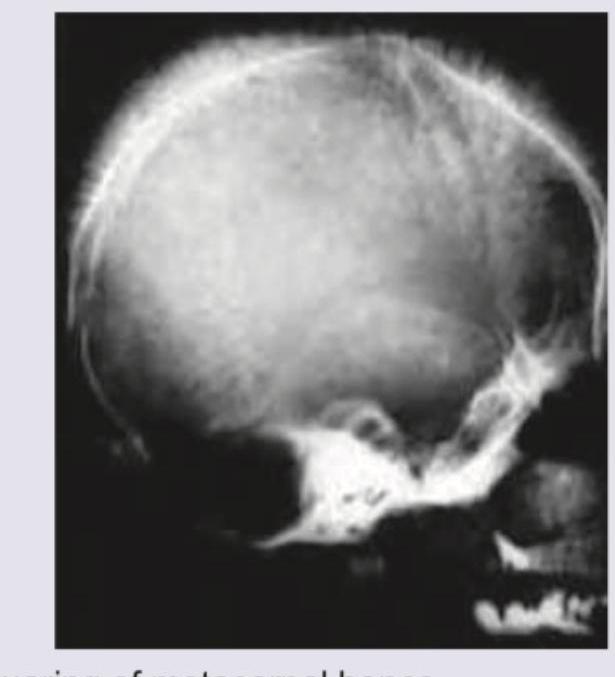
An 8-year-old boy with requirement of multiple blood transfusions. X-ray skull was performed. All are true about the condition shown except:

A 12-year-old child presents with acute kidney injury after a bout of dysentery. Not seen is:
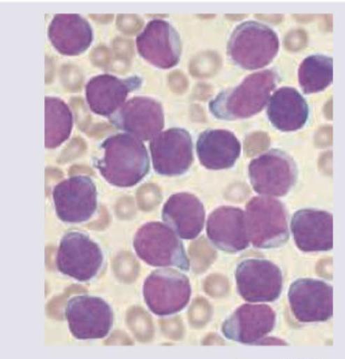
A 7-year-old boy presents with fever and weight loss. On examination he had pallor with lymphadenopathy. Peripheral smear is shown below. Diagnosis is: (AIIMS May 2017)

A 10-year-old boy presented with fatigue. Investigations revealed: Hemoglobin, 9 g/dL; MCV, 60 fL; MCH, 20 pg; and serum ferritin, 185 µg/L. The TLC was elevated and showed predominant lymphocytes and neutrophils. What is the likely diagnosis in this patient? **Normal values:** - Serum ferritin: 50-150 µg/L
Which of the following does not require a lumbar puncture in children?
A previously healthy child has sudden onset of red spots on body. There is a history of a preceding viral infection 1-4 weeks before the onset.
Normal reticulocyte count at birth is
Which of the following is the most common hematologic malignancy associated with Neurofibromatosis-1 (NF-1) in a child?
False about Shwachman-Diamond syndrome
Which of the following is most specific for congenital Rubella syndrome?
Explanation: ***Iron chelation leads to bronze diabetes*** - **Bronze diabetes** is caused by **hemochromatosis** (iron overload), where excess iron deposits in the pancreas, leading to diabetes and skin bronzing. Iron chelation therapy is given to **prevent** or treat iron overload, therefore it would logically **prevent** or reverse bronze diabetes, not cause it. - While chronic blood transfusions can cause iron overload, leading to bronze diabetes, chelation therapy is the treatment to prevent this complication and associated organ damage. *Squaring of metacarpal bones* - This is a typical radiological finding in **thalassemia major**, caused by **expanded bone marrow** in response to chronic anemia. - The abnormal erythropoiesis leads to widening of the medullary cavity and thinning of the cortical bone, giving the metacarpals a characteristic rectangular or "squared" appearance. *Erythroid hyperplasia in liver and spleen* - In conditions like thalassemia, the body attempts to compensate for ineffective erythropoiesis by activating **extramedullary hematopoiesis** in organs like the liver and spleen. - This results in the proliferation of erythroid precursors (erythroid hyperplasia) in these organs, contributing to hepatosplenomegaly. *Splenectomy if transfusion requirement increases by 50% over last year* - **Splenectomy** is a consideration in patients with thalassemia who have significantly increased transfusion requirements, usually defined as a 30-50% increase, due to **hypersplenism**. - Hypersplenism leads to increased destruction of red blood cells and premature removal of transfused cells, necessitating frequent transfusions.
Explanation: ***Normal serum haptoglobin*** - In hemolytic conditions like HUS (which classically follows dysentery in children), **haptoglobin** is consumed as it binds to free hemoglobin, leading to **decreased serum haptoglobin levels**, not normal levels. - The patient's presentation of acute kidney injury after dysentery is highly suggestive of **hemolytic uremic syndrome (HUS)**, a form of microangiopathic hemolytic anemia where red blood cells are destroyed. *MAHA* - **Microangiopathic hemolytic anemia (MAHA)** is central to the pathophysiology of HUS, where red blood cells are mechanically fragmented as they pass through narrowed and damaged microvessels. - The presence of **schistocytes** on a peripheral blood smear is a hallmark of MAHA. *Schistocytes* - **Schistocytes** (fragmented red blood cells) are a key laboratory finding in HUS, resulting from the shearing of red blood cells as they navigate obstructed microvasculature. - Their presence confirms the diagnosis of **microangiopathic hemolytic anemia**. *Thrombocytopenia* - **Thrombocytopenia** is a common feature of HUS, caused by the consumption of platelets within the microthrombi that form in the damaged microcirculation. - This platelet consumption contributes to the characteristic triad of HUS: hemolytic anemia, thrombocytopenia, and acute kidney injury.
Explanation: ***ALL*** - The peripheral smear shows numerous **blasts** with **scanty cytoplasm**, **prominent nucleoli**, and a **high nuclear-to-cytoplasmic ratio**, which is characteristic of **acute lymphoblastic leukemia (ALL)**. - Clinical features such as **fever**, **weight loss**, **pallor**, and **lymphadenopathy** in a 7-year-old child are classic presentations of ALL, the most common childhood leukemia. *AML* - While AML also presents with acute symptoms and blasts, the blasts in AML typically have **more abundant cytoplasm**, and may contain **Auer rods**, which are not clearly visible here. - The **morphology of the blasts** in the image, with their uniformly high N/C ratio and immature appearance, points away from typical AML. *Aplastic anemia* - Aplastic anemia is characterized by **pancytopenia** in the peripheral blood and **hypocellular bone marrow**, meaning a significant reduction in all blood cell lines, and **lacks the presence of blasts**. - The image clearly displays a proliferation of immature cells (blasts), which is contrary to the pathology of aplastic anemia. *Juvenile myelomonocytic leukemia* - JMML is a rare disorder characterized by **monocytosis**, **splenomegaly**, and typically presents in very young children (median age 2 years), often with **rashes** and **adenopathy**. - While there is some overlap in symptoms, the predominant cell type in the smear does not suggest a significant monocytic component, and the clinical picture in a 7-year-old child is less typical for JMML without other defining features.
Explanation: ***Beta thalassemia trait*** - The combination of **microcytic hypochromic anemia** (low MCV, MCH) with **normal to elevated ferritin** (185 µg/L is above the normal range of 50-150 µg/L) is highly suggestive of **beta thalassemia trait**, as iron stores are typically adequate or increased. - An elevated TLC with predominant lymphocytes and neutrophils is nonspecific but does not rule out thalassemia, as secondary infections or inflammatory processes can occur. *Anemia of chronic disease* - While anemia of chronic disease can cause **microcytic or normocytic anemia** and elevated ferritin (as ferritin is an acute phase reactant), the degree of **microcytosis** (MCV 60 fL) is more profound than typically seen in ACD. - ACD usually involves **inflammation or infection**, but the lab values provided more strongly point away from pure ACD. *Iron deficiency anemia* - **Iron deficiency anemia** is characterized by **microcytic hypochromic anemia** but would present with **low serum ferritin** levels, indicating depleted iron stores. - The patient's **elevated ferritin** (185 µg/L) rules out iron deficiency as the primary cause. *Lead poisoning* - **Lead poisoning** can cause **microcytic anemia** by interfering with heme synthesis, but it is typically associated with a **basophilic stippling** on peripheral blood smear and elevated blood lead levels. - It does not typically present with **elevated ferritin** as a classic feature, and the overall clinical picture is more consistent with a genetic hemoglobinopathy.
Explanation: ***HL*** - While central nervous system (CNS) involvement is possible in Hodgkin lymphoma (HL), it is **rare** and does not routinely warrant a **lumbar puncture** for initial staging or surveillance in asymptomatic children. - HL primarily affects **lymph nodes** and the **spleen**, with CNS spread being an uncommon complication that typically presents with specific neurological symptoms. *AML* - **Acute myeloid leukemia (AML)** has a significant risk of **CNS involvement**, requiring a **lumbar puncture** for diagnostic staging and administration of intrathecal chemotherapy. - CNS prophylaxis and treatment are crucial in AML to prevent and manage **leptomeningeal disease**. *NHL* - **Non-Hodgkin lymphoma (NHL)**, particularly aggressive subtypes like Burkitt lymphoma or lymphoblastic lymphoma, has a **high propensity for CNS spread**. - A **lumbar puncture** is essential for staging to detect CNS involvement and guide the need for intrathecal chemotherapy or radiation. *ALL* - **Acute lymphoblastic leukemia (ALL)** carries a well-documented **high risk of CNS infiltration**, necessitating routine **lumbar punctures** at diagnosis for CNS staging and throughout treatment for intrathecal chemotherapy. - CNS prophylaxis is a cornerstone of ALL treatment to prevent **leptomeningeal relapse**.
Explanation: ***Idiopathic thrombocytopenic purpura (ITP)*** - This presentation, especially in a previously healthy child with a preceding viral infection 1-4 weeks prior, is highly characteristic of **acute ITP**, leading to **purpuric rash** (red spots). - The preceding viral infection often triggers an autoimmune response causing destruction of **platelets**, resulting in **thrombocytopenia**. *Dengue fever* - Dengue fever typically presents with **acute onset of fever**, **headache**, **myalgia**, and a rash that appears 3-4 days after fever onset, often with a shorter incubation period than 1-4 weeks. - While it can cause petechiae due to **thrombocytopenia**, the symptom constellation does not perfectly align with the scenario, particularly the sudden onset of spots without mention of fever or other acute symptoms. *Hemophilia A* - **Hemophilia A** is a **hereditary bleeding disorder** causing deficits in **Factor VIII**, leading to spontaneous bleeding into joints and muscles, and prolonged bleeding after trauma. - It does not present as sudden onset red spots (petechiae/purpura) following a viral infection but rather as larger **hematomas** or **hemarthroses**, and it's a chronic condition, not typically triggered by recent infection. *Thrombotic thrombocytopenic purpura (TTP)* - TTP is characterized by the **pentad of symptoms**: **fever**, **neurological symptoms**, **renal dysfunction**, **microangiopathic hemolytic anemia**, and **thrombocytopenia**. - While it involves thrombocytopenia and can cause purpura, the patient's presentation lacks the other severe systemic features typically associated with TTP, and it's less commonly triggered by a simple viral infection in children.
Explanation: ***2-6%*** - At birth, the normal **reticulocyte count** is elevated primarily due to the physiological stress of **adapting to extrauterine life** and increased erythropoiesis. - This higher range reflects the body's need for a rapid turnover of red blood cells to meet oxygen demands after birth. *1-2%* - This range is considered a **normal reticulocyte count** for **older children and adults**, indicating a steady state of red blood cell production. - It does not account for the **physiological erythropoietic surge** observed in healthy neonates. *6-10%* - While higher than adult levels, a range of **6-10%** in a neonate would still be considered unusually high and might suggest **pathological hemolysis** or significant **blood loss**. - Without other supporting clinical signs, this level is typically higher than the **physiological norm**. *30-40%* - A reticulocyte count of **30-40%** at birth is **extremely elevated** and is highly indicative of a severe underlying condition such as **hemolytic disease of the newborn** or significant **neonatal hemorrhage**. - This level is far beyond the **normal physiological response** and requires urgent investigation.
Explanation: ***Juvenile Myelomonocytic Leukemia (JMML)*** - **JMML** is a myelodysplastic/myeloproliferative neoplasm that is strongly associated with **NF-1** in children, particularly due to mutations in the *NF1* gene. - Children with **NF-1** have a significantly increased risk of developing **JMML** compared to the general pediatric population. *Chronic Myeloid Leukemia (CML)* - While CML can occur in children, it is typically associated with the **Philadelphia chromosome (BCR-ABL1 fusion gene)** and not a common tumor directly linked to NF-1. - **CML** is relatively rare in childhood compared to other leukemias and is not a characteristic complication of NF-1. *Acute Lymphoblastic Leukemia (ALL)* - **ALL** is the most common childhood cancer overall, but its association with **NF-1** is not as specific or strong as that of **JMML**. - While children with NF-1 may have a slightly increased risk of certain cancers, **ALL** is not the *most common* tumor directly linked to NF-1. *Acute Myeloid Leukemia (AML)* - **AML** has a weak association with **NF-1**, particularly specific subtypes, but it is less frequent and less specifically linked than **JMML**. - The direct genetic pathway involving the **NF1 gene** mutation in the development of **AML** is not as clearly defined as it is for **JMML**.
Explanation: ***Leucocytosis*** - **Leucocytosis** (an increase in white blood cells) is generally **not** a feature of Shwachman-Diamond syndrome (SDS); rather, patients typically experience **neutropenia** (low neutrophils) due to bone marrow dysfunction. - This persistent or intermittent neutropenia is a hallmark of the immune deficiency seen in SDS, making leucocytosis an incorrect finding. *Bone marrow dysfunction* - **Bone marrow dysfunction** is a defining characteristic of Shwachman-Diamond syndrome, leading to various **cytopenias**, most notably **neutropenia**. - This dysfunction can also manifest as anemia or thrombocytopenia, contributing to the overall morbidity of the disease. *Exocrine pancreatic insufficiency* - **Exocrine pancreatic insufficiency** is a primary clinical feature of Shwachman-Diamond syndrome, leading to **malabsorption** and **failure to thrive**. - This insufficiency is due to abnormal pancreatic development and is distinct from the more severe pancreatic involvement seen in cystic fibrosis. *Short stature* - **Short stature** is a common finding in children with Shwachman-Diamond syndrome, often resulting from a combination of **growth plate abnormalities** and **malnutrition** due to pancreatic insufficiency. - It is considered a key **skeletal manifestation** of the disease, along with metaphyseal chondrodysplasia.
Explanation: ***Triad of CRS are cataract, cardiac defects, sensorineural deafness*** - The **classic Gregg triad** of **cataracts**, **cardiac defects** (especially patent ductus arteriosus and pulmonary artery stenosis), and **sensorineural deafness** is the **most specific and pathognomonic** feature of **congenital Rubella syndrome**. - While individual components can occur in other conditions, the **combination of this triad** is highly specific for CRS and distinguishes it from other congenital infections. - This triad was first described by **Norman Gregg** in 1941 and remains the hallmark diagnostic feature of congenital rubella syndrome. *Blueberry muffin rash is seen* - The **blueberry muffin rash** (dermal erythropoiesis) presents as purpuric lesions or small dark blue papules and can be seen in congenital rubella syndrome. - However, this finding is **NOT specific to rubella** and occurs in multiple congenital infections including **CMV, toxoplasmosis, parvovirus B19**, and can also be seen in neonatal malignancies like neuroblastoma. - While characteristic, it is less specific than the Gregg triad for diagnosing CRS. *Infection is most serious in the first trimester of pregnancy* - Maternal rubella infection during the **first trimester** carries the highest risk (up to 85% if infected before 12 weeks) of severe multi-organ abnormalities due to rapid organogenesis. - While true, this describes the **timing and severity** of infection rather than a specific clinical feature that distinguishes rubella from other congenital infections. - Many congenital infections (CMV, toxoplasmosis, HSV) are also more severe when acquired in early pregnancy. *Virus can be isolated up to 12 months after birth* - Infants with **congenital Rubella syndrome** can shed virus in bodily fluids (urine, nasopharyngeal secretions) for **12 months or longer** after birth. - This prolonged viral shedding is important for **infection control** and isolation precautions but is a virological characteristic rather than a specific diagnostic clinical feature. - Other congenital infections (CMV) can also demonstrate prolonged viral shedding in infants.
Anemias in Children
Practice Questions
Hemoglobinopathies
Practice Questions
Hemolytic Anemias
Practice Questions
Nutritional Anemias
Practice Questions
Thrombocytopenia
Practice Questions
Bleeding Disorders
Practice Questions
Thrombotic Disorders
Practice Questions
White Blood Cell Disorders
Practice Questions
Bone Marrow Failure Syndromes
Practice Questions
Blood Component Therapy
Practice Questions
Hemophilia and Von Willebrand Disease
Practice Questions
Evaluation of Bleeding Tendencies
Practice Questions
Get full access to all questions, explanations, and performance tracking.
Start For Free