Which of the following is NOT true regarding fetal red blood cells?
A 7-year-old child is brought to the clinic with complaints of fatigue and poor concentration. The mother reports that the child has been eating non-food items such as chalk and soil for the past few months. A peripheral blood smear image shows microcytic, hypochromic red blood cells. Which of the following is the most likely diagnosis?
A child with progressive pallor and bone pain has an elevated HbS based on the HPLC report. Which is the best treatment to manage hemolysis in this patient?
A young boy presented with petechiae and his platelet count was 10,000/cu mm. Bone marrow aspirate revealed normal cellularity with megakaryocyte hyperplasia. Most appropriate initial therapy?
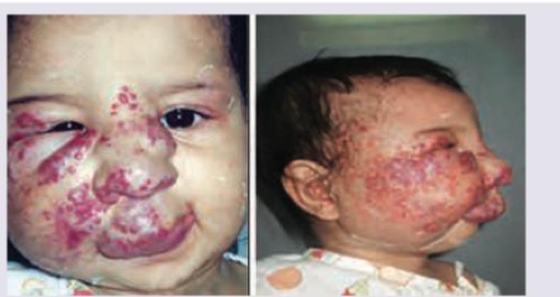
A 1-year-old infant with the following lesion on face. CNS examination was normal. Blood counts show thrombocytopenia with P. smear suggestive of microangiopathic changes. Probable diagnosis is:

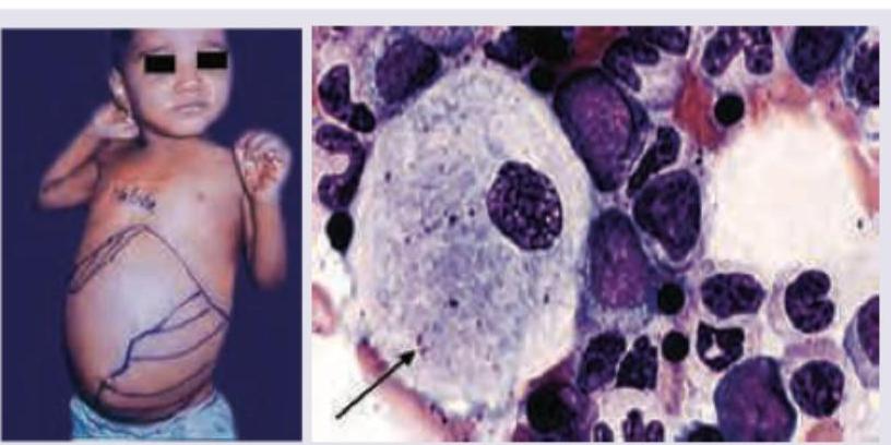
A 2-year-old child presents with growth retardation, pallor, bruising and has palpable spleen 5 cm below left costal margin. Bone marrow examination is shown below. Which is incorrect about the clinical diagnosis? (Recent NEET Pattern 2016-17)

A 3-year-old child presents with bleeding from nose and Periorbital Ecchymosis. Sternal tenderness and bone pain is present. Peripheral smear shows presence of fragmented RBC and helmet cells. The most probable diagnosis is? (Recent NEET Pattern 2016-17)
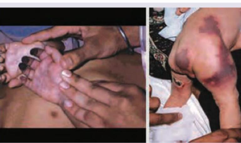
A 6-month-old child brought by parents for diffuse ecchymosis on extremities and trunk. Probable diagnosis is? (Recent NEET Pattern 2016-17)

A 3-year-old child presents with sudden onset generalized petechiae and bruise on forehead. Sternal tenderness is absent and liver and spleen are not palpable. Bone marrow aspiration is normal. Probable cause is? (Recent NEET Pattern 2016-17)
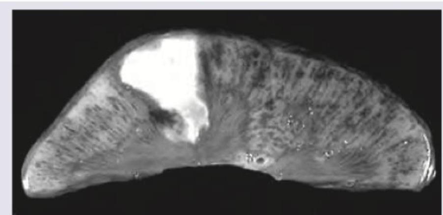
A 10-year-old Sindhi boy presents with recurrent episodes of bone pain. Specimen shows:

Explanation: **Explanation:** **1. Why Option A is Correct (The Underlying Concept):** Fetal red blood cells (RBCs) are characterized by **low levels of 2,3-Diphosphoglycerate (2,3-DPG)**. Furthermore, Fetal Hemoglobin (HbF) has a structurally lower affinity for 2,3-DPG compared to adult hemoglobin (HbA). Since 2,3-DPG normally functions to stabilize the "T" (tense) state of hemoglobin and promote oxygen unloading, its low levels/reduced binding in fetuses result in a **leftward shift** of the oxygen-dissociation curve. This allows the fetus to maintain a high affinity for oxygen, facilitating the extraction of oxygen from maternal blood across the placenta. **2. Analysis of Incorrect Options:** * **B. Decreased carbonic anhydrase activity:** This is a **true** characteristic. Fetal RBCs have significantly lower levels of carbonic anhydrase, which can lead to a slower conversion of CO2 to bicarbonate, though this is physiologically compensated in utero. * **C. Decreased lifespan:** This is **true**. Fetal RBCs have a shorter survival time (approximately **60–90 days**) compared to the 120-day lifespan of adult RBCs. This contributes to the physiological jaundice often seen in neonates. * **D. High RBC volume:** This is **true**. Neonatal RBCs are **macrocytic** (High MCV), typically ranging from 100–120 fL at birth. **3. NEET-PG High-Yield Pearls:** * **HbF Structure:** Composed of two alpha and two **gamma** chains ($\alpha_2\gamma_2$). * **P50 Value:** The P50 (partial pressure of O2 at which 50% of Hb is saturated) is **lower** in fetuses (~19 mmHg) compared to adults (~27 mmHg), reflecting higher affinity. * **Metabolism:** Fetal RBCs have higher activity of enzymes in the glycolytic pathway (like hexokinase) but lower activity of others (like phosphofructokinase).
Explanation: ***Iron deficiency anemia*** - The patient's history of **pica** (craving and eating non-food items like chalk and soil) is a classic clinical sign of iron deficiency. - The peripheral blood smear confirms this diagnosis by showing **microcytic** (small) and **hypochromic** (pale) red blood cells, which result from impaired hemoglobin synthesis due to a lack of iron. *Vitamin B12 deficiency* - This condition causes **macrocytic anemia**, where red blood cells are larger than normal (high MCV), which is the opposite of the findings in this case. - Peripheral smear findings in B12 deficiency typically include **macro-ovalocytes** and **hypersegmented neutrophils**, neither of which is described or shown. *Thalassemia major* - Although thalassemia causes a **microcytic, hypochromic anemia**, it is a genetic disorder that typically presents in infancy with severe symptoms like failure to thrive and massive **hepatosplenomegaly**. - Pica is not a characteristic feature of thalassemia; it is strongly associated with iron deficiency anemia. *Lead poisoning* - Lead poisoning can cause microcytic anemia, but a key finding on the peripheral smear is **basophilic stippling**, which is not the prominent feature here. - While pica can be a risk factor for lead ingestion, pica itself is a more direct and classic symptom of underlying **iron deficiency**.
Explanation: ***Voxelotor*** - It is a **hemoglobin polymerization inhibitor** that stabilizes the oxygenated form of red blood cells, preventing **sickling** and subsequent hemolytic anemia, thereby directly managing the hemolysis. - It significantly improves **hemoglobin levels** and reduces markers of hemolysis, such as **indirect bilirubin** and **reticulocyte count**. *Hydroxyurea* - Its primary mechanism is inducing the production of **fetal hemoglobin (HbF)**, thereby diluting the concentration of HbS and *indirectly* reducing hemolysis over time. - Although crucial for managing **vaso-occlusive crises** (VOCs), it is not as direct an anti-hemolytic agent as Voxelotor. *Azacytidine* - This is a **DNA methyltransferase inhibitor**, primarily used in high-risk **Myelodysplastic Syndrome (MDS)** and Acute Myeloid Leukemia (AML). - While it can induce HbF like Hydroxyurea, it is not a standard or approved frontline treatment for the routine management of Sickle Cell Disease (SCD). *Bortezomib* - This drug is a **proteasome inhibitor** used exclusively in the treatment of **Multiple Myeloma** and certain related plasma cell dyscrasias. - It has no therapeutic role or clinical indication in the specific management of hemolysis or the underlying pathophysiology of SCD.
Explanation: ***IVIG*** - The presentation (young patient, **petechiae**, extremely low platelet count of 10,000/cu mm, and **megakaryocyte hyperplasia** in bone marrow) is classic for **Immune Thrombocytopenia (ITP)**. - Given the severe thrombocytopenia (platelets <20,000/cu mm) and active bleeding risk (petechiae), the initial treatment of choice is typically **IVIG (Intravenous Immunoglobulin)** or corticosteroids to rapidly increase the platelet count. *Aspirin* - **Aspirin** is an antiplatelet agent that **inhibits platelet aggregation**; it is strictly contraindicated in severe thrombocytopenia like ITP as it would increase the risk of hemorrhage. - It is used for pain relief or secondary prevention of cardiovascular events, not for treating low platelet counts. *Bone Marrow Transplant* - **Bone marrow transplant** is a radical and highly risky procedure reserved for refractory, life-threatening hematological malignancies or severe aplastic anemia, or failed therapy for ITP. - It is never the appropriate **initial therapy** for acute ITP, which is predominantly an autoimmune disorder of peripheral platelet destruction. *Methotrexate* - **Methotrexate** is an immunosuppressive drug used typically in chronic inflammatory conditions (like rheumatoid arthritis) or malignancies. - It is considered a second or third-line treatment option, or part of a more aggressive immunosuppression regimen, for ITP that has failed initial therapy (steroids/IVIG), not the immediate first choice.
Explanation: ***Kasabach-Merritt syndrome*** - The image shows a large **vascular lesion (hemangioma)** on the infant's face, consistent with a rapidly enlarging vascular tumor. - The presence of **thrombocytopenia** and **microangiopathic hemolytic anemia** (suggested by microangiopathic changes on peripheral smear) in conjunction with a vascular lesion are hallmarks of Kasabach-Merritt phenomenon, a life-threatening complication associated with certain rapidly growing vascular tumors like kaposiform hemangioendothelioma or tufted angioma. *Tuberous sclerosis* - Tuberous sclerosis involves various organs and its cutaneous manifestations primarily include **ash-leaf spots**, **facial angiofibromas** (which typically appear later in childhood/adolescence), and shagreen patches. - It is a **neurocutaneous syndrome** characterized by CNS abnormalities, such as subependymal nodules and cortical tubers, which are explicitly stated as normal in this case. *Thrombotic thrombocytopenic purpura* - TTP is characterized by the pentad of **thrombocytopenia**, **microangiopathic hemolytic anemia**, fever, renal failure, and neurological symptoms, but it is typically an acquired disorder in adults, rarely seen in infants, and not associated with a visible large vascular tumor as seen in the image. - The **absence of significant CNS involvement** in this infant makes TTP less likely, and the primary presentation with a large facial lesion points away from TTP as the underlying cause. *Sturge-Weber syndrome* - Sturge-Weber syndrome is characterized by a **port-wine birthmark (nevus flammeus)**, usually unilateral, in the distribution of the trigeminal nerve, and is associated with **leptomeningeal angioma** leading to seizures and neurological deficits. - While it includes a prominent facial vascular lesion, the description of normal CNS examination directly contradicts a key diagnostic criterion for Sturge-Weber syndrome, and the image does not clearly depict a typical port-wine stain.
Explanation: ***Defect on chromosome 3*** - The clinical presentation (growth retardation, pallor, bruising, massive splenomegaly) and the bone marrow image showing a **Gaucher cell** (macrophage with a characteristic "crinkled paper" or "crumpled tissue paper" cytoplasm due to glucocerebroside accumulation) are classic for **Gaucher disease**. - Gaucher disease is caused by a **deficiency of the enzyme glucocerebrosidase**, which is encoded by the GBA gene located on **chromosome 1q21**, not chromosome 3. Therefore, a defect on chromosome 3 is incorrect. *Glucosylceramide accumulation in reticulo-endothelial cells* - This is a correct feature of Gaucher disease, where the deficient enzyme **glucocerebrosidase** leads to the accumulation of its substrate, **glucocerebroside (glucosylceramide)**, primarily within macrophages (reticulo-endothelial cells). - These lipid-laden macrophages are known as **Gaucher cells** and are pathognomonic for the disease. *Bleeding episodes* - **Thrombocytopenia** (low platelet count) is a common hematological manifestation in Gaucher disease, often due to **hypersplenism** caused by massive splenomegaly and direct bone marrow infiltration. - This low platelet count can lead to an increased tendency for **bruising and bleeding episodes**, which is consistent with the patient's bruising. *Erlenmeyer flask deformity of the distal femur* - This is a characteristic **radiological finding** in Gaucher disease, particularly in the long bones. - It refers to the **widening of the distal metaphysis** of the femur, resembling an Erlenmeyer flask, due to bone marrow infiltration by Gaucher cells.
Explanation: ***Acute myeloid Leukemia*** - The combination of **bleeding manifestations** (nose bleeding, periorbital ecchymosis), **bone pain and sternal tenderness** (from marrow infiltration), and **fragmented RBCs with helmet cells** (schistocytes) on peripheral smear is highly suggestive of **Acute Myeloid Leukemia complicated by DIC**. - **Fragmented RBCs and helmet cells** indicate **microangiopathic hemolytic anemia (MAHA)**, which in the context of acute leukemia suggests **disseminated intravascular coagulation (DIC)** - a well-known complication of AML, especially **acute promyelocytic leukemia (AML-M3)**. - **Bone pain and sternal tenderness** are characteristic features of leukemic infiltration of bone marrow. - **Bleeding** occurs due to thrombocytopenia from marrow replacement and consumption in DIC. *Battered baby syndrome* - While trauma/abuse can cause periorbital ecchymosis and bleeding, the presence of **sternal tenderness, bone pain** (suggesting marrow pathology), and **schistocytes on peripheral smear** is NOT typical of trauma alone. - Schistocytes would only occur in trauma if there is severe DIC from massive injury with shock, which would present as a critically ill child with multiorgan dysfunction. - The constellation of findings here points to **hematologic malignancy**, not physical abuse. *Acute Idiopathic thrombocytopenic purpura* - ITP causes **bleeding manifestations** due to isolated **thrombocytopenia**, leading to nosebleeds and ecchymosis. - However, **bone pain, sternal tenderness**, and **fragmented RBCs with helmet cells** are NOT features of ITP. - ITP has a normal peripheral smear except for decreased platelets. *Hemophilia A* - Hemophilia A is a **coagulation disorder** characterized by **factor VIII deficiency**, leading to bleeding into joints and deep tissues. - **Bone pain and sternal tenderness** are not characteristic of hemophilia. - **Fragmented RBCs and helmet cells** do not occur in hemophilia. - Periorbital ecchymosis from minor trauma can occur, but the complete clinical picture does not fit.
Explanation: ***Protein C deficiency*** - A 6-month-old with **diffuse ecchymoses on extremities and trunk** is classic for **homozygous protein C deficiency** presenting as **purpura fulminans**. - Severe congenital protein C deficiency typically manifests in **early infancy** (within first few weeks to months) with widespread purpuric lesions, skin necrosis, and thrombotic complications. - The presentation includes **large ecchymotic patches** that can progress to necrosis, affecting extremities and trunk as described. - This is the **most likely diagnosis** for this age group and clinical presentation in the absence of other systemic features. *Disseminated intravascular coagulation* - DIC causes diffuse bleeding and purpura due to consumption coagulopathy and microthrombi formation. - However, DIC is a **secondary complication** requiring an underlying trigger (sepsis, trauma, malignancy) with acute systemic illness. - The child would typically present with **multiple bleeding sites**, systemic signs, and evidence of the underlying condition—not isolated ecchymosis. *A.P.L.A.S (Antiphospholipid Antibody Syndrome)* - Neonatal antiphospholipid syndrome from maternal antibodies presents in the **neonatal period** (first 28 days of life), not at 6 months. - APLAS primarily causes **thrombotic events** (stroke, renal vein thrombosis) rather than diffuse ecchymosis alone. - This diagnosis is **extremely rare** in infancy and would not be the probable diagnosis for isolated ecchymotic presentation at this age. *Dengue hemorrhagic fever* - Dengue requires **exposure in endemic areas** and presents with fever, headache, myalgia, and characteristic rash followed by hemorrhagic manifestations. - More common in **older children and adults**; the 6-month-old age and absence of fever/systemic symptoms make this unlikely. - The clinical picture describes ecchymosis without mention of fever or other dengue features.
Explanation: ***Acute ITP*** - A 3-year-old child presenting with **sudden onset petechiae and ecchymoses (bruise on forehead)**, without signs of systemic illness (no sternal tenderness, non-palpable liver/spleen), and a normal bone marrow is highly suggestive of **Idiopathic Thrombocytopenic Purpura (ITP)**. ITP is characterized by isolated thrombocytopenia due to autoimmune destruction of platelets. - The **normal bone marrow**, in particular, rules out conditions like leukemia and aplastic anemia, where bone marrow pathology would be evident. *Aplastic anemia* - **Aplastic anemia** would typically present with **pancytopenia** (affecting all blood cell lines), leading to symptoms like pallor, fatigue, infections, and bleeding. - A **bone marrow aspiration** in aplastic anemia would show a **hypocellular marrow** with a reduction in hematopoietic cells, which contradicts the "normal" finding in this case. *Acute leukemia* - **Acute leukemia** in children often presents with **bone pain (sternal tenderness)**, fever, fatigue, and organomegaly (enlarged liver and spleen) due to infiltration of leukemic cells. - A **bone marrow aspiration** would reveal a **hypercellular marrow** with a large proportion of blasts, which is not consistent with the "normal" bone marrow finding provided. *Dengue hemorrhagic fever* - **Dengue hemorrhagic fever** typically presents with symptoms like high fever, severe headache, muscle and joint pain, and often signs of bleeding (like petechiae and bruising) and can lead to severe thrombocytopenia. - However, it is usually preceded by a **fever** and other systemic symptoms, which are not mentioned in this sudden onset presentation in the given case scenario.
Explanation: ***Spleen infarction*** - The image shows a **wedge-shaped, pale area** within the spleen, consistent with an **infarct**. Splenic infarctions are common in conditions leading to **vaso-occlusion**, such as **sickle cell disease**. - A 10-year-old Sindhi boy with **recurrent bone pain** suggests a **hemoglobinopathy**, particularly sickle cell disease (or a related condition like **thalassemia-sickle beta**), where red blood cells can obstruct splenic blood vessels leading to infarction. *Liver hematoma* - A **hematoma** is a collection of blood, often caused by trauma or hemorrhage. The image depicts a **necrotic area** (infarct) rather than a blood clot within the liver. - While liver issues can occur in some hematologic disorders, the characteristic wedge shape points away from a simple hematoma. *Heart infarction* - A **heart infarction** (myocardial infarction) affects the heart muscle. The specimen in the image is clearly an **abdominal organ**, identifiable as the spleen due to its characteristic shape and internal architecture. - The clinical presentation of bone pain is not directly indicative of a heart infarction. *Adrenal hemorrhage* - **Adrenal hemorrhage** would involve bleeding within the adrenal glands, which are small glands located on top of the kidneys. The organ shown in the image is much larger and shaped like a spleen. - Bone pain is not a typical symptom of adrenal hemorrhage.
Anemias in Children
Practice Questions
Hemoglobinopathies
Practice Questions
Hemolytic Anemias
Practice Questions
Nutritional Anemias
Practice Questions
Thrombocytopenia
Practice Questions
Bleeding Disorders
Practice Questions
Thrombotic Disorders
Practice Questions
White Blood Cell Disorders
Practice Questions
Bone Marrow Failure Syndromes
Practice Questions
Blood Component Therapy
Practice Questions
Hemophilia and Von Willebrand Disease
Practice Questions
Evaluation of Bleeding Tendencies
Practice Questions
Get full access to all questions, explanations, and performance tracking.
Start For Free