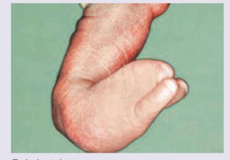
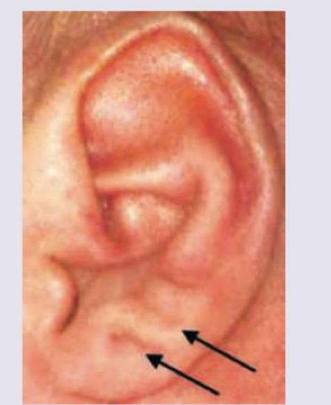
The image shows presence of:

A child suffering from respiratory and feeding difficulties was brought for consultation. All of the following are to be considered in differential diagnosis of the child except:
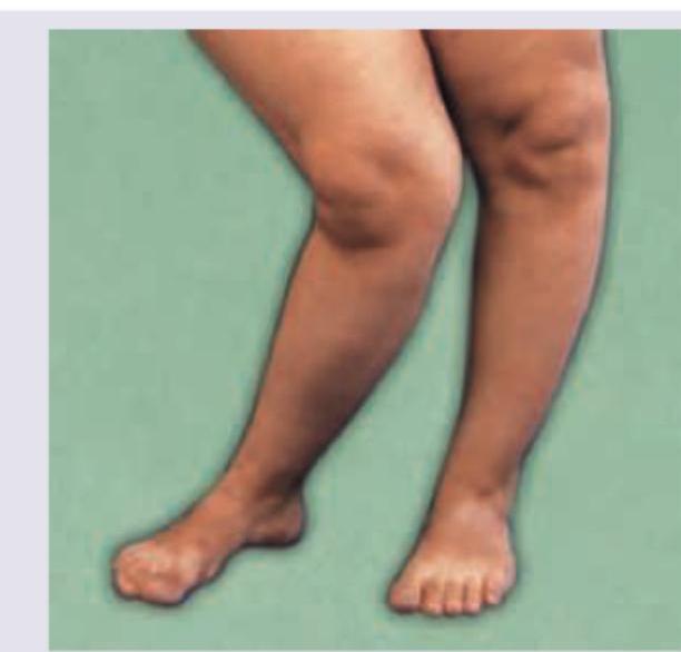
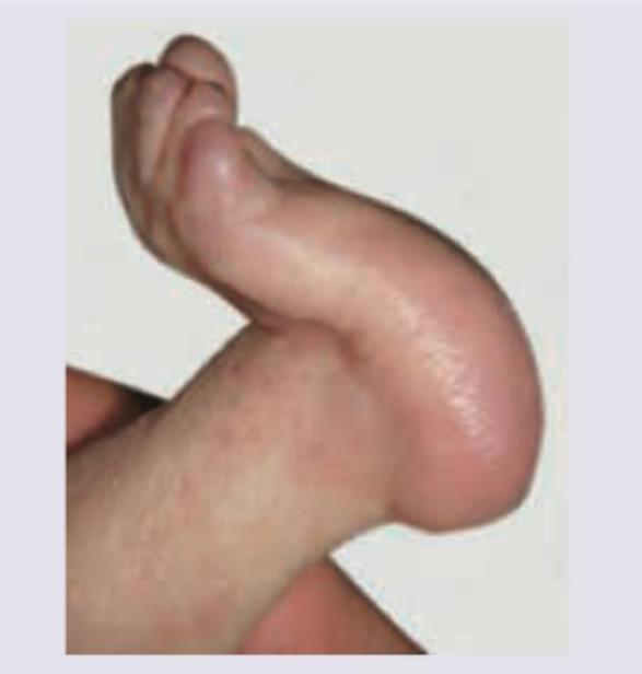
Identify the deformity seen in this child with rickets:

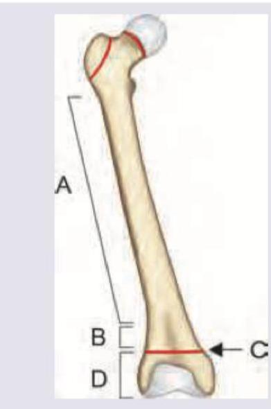
In a child with rickets which part of the bone is affected?

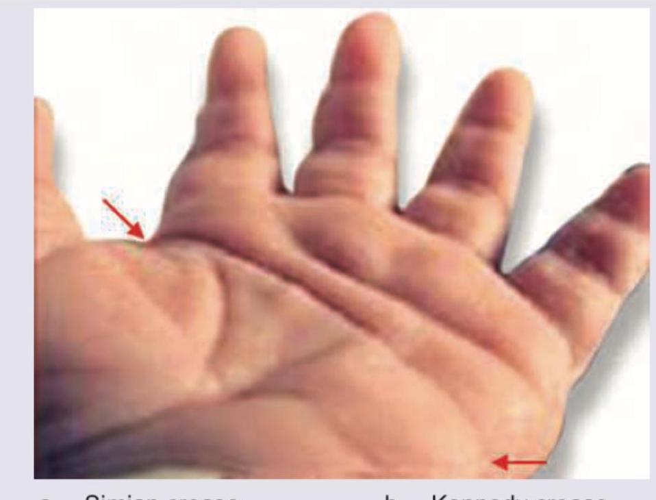
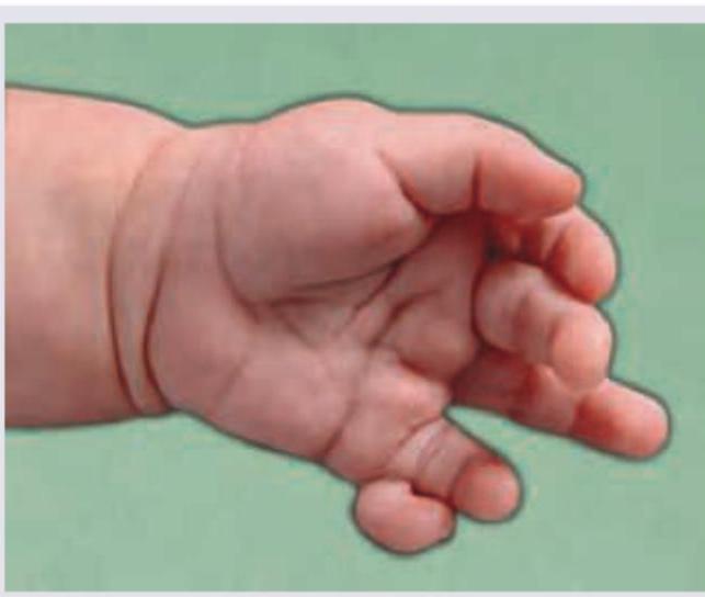
During evaluation of a child with Down syndrome, the following finding is noted. Identify?

Which of the following is associated with this condition?

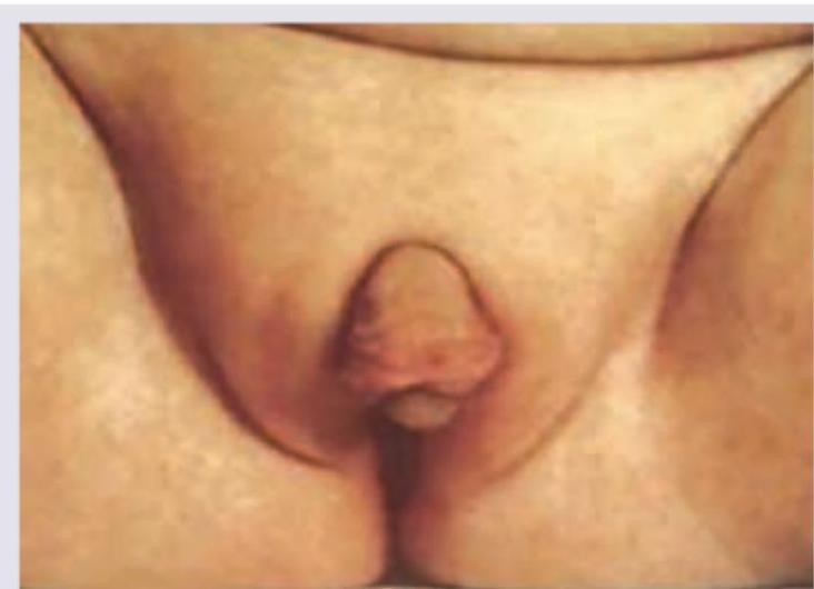
During evaluation of a child with obesity, following finding is observed. What is the association?

This baby has obesity along with organomegaly. Comment on the diagnosis from the hint given in the image?

Which is true about an infant with failure to thrive and the following findings?

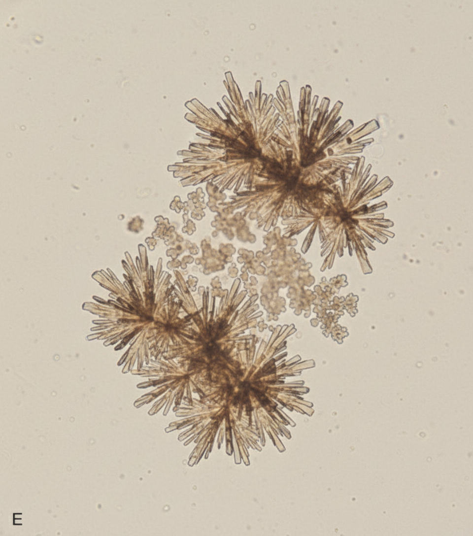
A 6-year-old child presents with recurrent urinary tract infections. Urine microscopy findings are shown in the image below. What type of crystalluria is depicted?

Explanation: ***Ectrodactyly*** - The image clearly shows a malformation of the foot where the **central digits are missing**, creating a characteristic **cleft** or "lobster claw" appearance. This is the defining feature of ectrodactyly (also called split hand/foot malformation). - Ectrodactyly can affect hands or feet and may occur as an **isolated anomaly** or as part of genetic syndromes like EEC syndrome (Ectrodactyly-Ectodermal dysplasia-Clefting). - Inheritance is typically **autosomal dominant** with variable expressivity. *Polydactyly* - Polydactyly refers to the presence of extra digits (supernumerary fingers or toes), which is not observed in the image. - The image shows a deficiency of central digits with a cleft, not an excess of digits. *Cutis aplasia* - Cutis aplasia (aplasia cutis congenita) is characterized by localized absence of skin at birth, most commonly affecting the scalp. - The image displays a limb malformation involving bone and soft tissue structure, not a skin defect. *TAR syndrome* - TAR (Thrombocytopenia-Absent Radius) syndrome is characterized by bilateral absence or hypoplasia of the radii in the forearms, along with thrombocytopenia. - While TAR involves limb malformations, the image shows a foot deformity (ectrodactyly), and there is no evidence of absent radii or hematological abnormalities visible in the image.
Explanation: ***Turner syndrome*** - Turner syndrome is a **chromosomal disorder** (45,XO) primarily affecting females, characterized by **short stature**, **gonadal dysgenesis**, and various physical features that typically do not include the severe craniofacial malformations causing immediate respiratory and feeding difficulties seen in the other syndromes. - While it can involve **cardiac anomalies** (e.g., coarctation of the aorta), these generally do not manifest as immediate life-threatening respiratory or feeding issues in the newborn period in the same way as disorders with significant craniofacial malformations. *Treacher Collins syndrome* - This is a **craniofacial dysostosis** characterized by **mandibulofacial anomalies**, including hypoplasia of the zygomatic bones and mandible, and downward-sloping palpebral fissures. - These malformations often lead to **airway obstruction** (due to micrognathia and glossoptosis) and **feeding difficulties**. *Pierre-Robin syndrome* - Characterized by the triad of **micrognathia**, **glossoptosis** (posterior displacement of the tongue), and **cleft palate**. - The combination of a small jaw and a tongue that falls back can cause severe **upper airway obstruction** and significant **feeding problems**. *Velocardiofacial syndrome* - Also known as **22q11.2 deletion syndrome**, this condition is associated with a wide range of features including **palatal abnormalities** (cleft palate or velopharyngeal insufficiency), **cardiac defects**, and distinctive facial features. - The palatal issues can lead to **feeding difficulties**, and cardiac or upper airway anomalies can contribute to **respiratory problems**.
Explanation: ***Genu varum (Bow legs)*** - The image shows the knees bowed outwards resulting in a **bow-legged appearance**, which is characteristic of **genu varum**. - **Rickets** is a common cause of genu varum, as the softened bones are unable to bear weight and bend outwards. *Genu valgum (knock knees)* - **Genu valgum** presents as the knees bending inwards, appearing to "knock" against each other, which is the opposite of what is seen in the image. - While rickets can also cause genu valgum, the depicted deformity clearly shows outward bowing. *Windswept deformity* - A **windswept deformity** involves one knee being in **valgus** (knock-kneed) and the other in **varus** (bow-legged). - The image does not show this combination; both knees appear to be bowed outwards. *Gunstock deformity* - **Gunstock deformity** is an angular deformity of the elbow, typically related to a **supracondylar humerus fracture**. - This deformity affects the upper limb and is not related to the lower limb presentation in the image.
Explanation: ***C*** - C points to the **growth plate (physis) and metaphyseal region**, which is the **primary site of pathology in rickets**. - Rickets causes defective mineralization at the growth plate where endochondral ossification occurs, leading to accumulation of unmineralized osteoid and disorganized chondrocyte columns. - The **metaphysis** (the region including the growth plate) shows the characteristic radiological findings: widening, fraying, cupping, and loss of sharp margins. *A* - A indicates the **diaphysis** (shaft of the bone), which consists of mature cortical bone. - The diaphysis is **not primarily affected** in rickets, though severe cases may lead to bowing deformities due to bone softening. - The initial and primary pathological changes occur at the actively growing regions, not the shaft. *B* - B points to the **metaphysis**, which is the flared region of long bone adjacent to the growth plate. - The metaphysis is indeed significantly affected in rickets, showing widening and irregular mineralization. - However, if the image distinguishes between the broader metaphyseal region (B) and the specific growth plate zone (C), then C represents the more precise site where the pathological process of defective mineralization originates. *D* - D points to the **epiphysis**, the rounded end of the long bone that forms part of the joint. - The epiphysis consists of already-formed bone and is **not the primary site** of rickets pathology. - Rickets affects the zone of provisional calcification at the growth plate, not the pre-existing epiphyseal bone.
Explanation: ***Simian crease*** - The image displays a **single palmar crease** (also known as a simian crease or transverse palmar crease), which is a common physical finding in individuals with **Down syndrome (Trisomy 21)**. - This crease runs straight across the palm, often replacing the usual two major palmar creases. *Kennedy crease* - This is **not a recognized medical term** for a palmar crease or any specific dermatological finding. - The feature shown in the image is a clearly defined, single transverse palmar crease. *Sydney line* - A **Sydney line** is another type of palmar crease abnormality where the proximal transverse crease extends across the entire palm, reaching the ulnar edge. - While it is a variation of palmar creases, the crease in the image appears as a single, bold transverse crease, more consistent with a **simian crease**. *Sandal gap* - A **sandal gap** refers to a wide space between the first and second toes, sometimes seen in newborns, including those with Down syndrome. - This finding relates to the **foot**, not the hand, and is not depicted in the provided image.
Explanation: ***Edward syndrome*** - The image shows **rocker-bottom feet**, which is a characteristic skeletal deformity seen in Edward syndrome (**Trisomy 18**). - Rocker-bottom feet present as a **convex sole** with prominent heel and a **vertical talus**, creating a "rocker" appearance, and is one of the classic **dysmorphic features** of Trisomy 18. - Other features of Edward syndrome include **overlapping fingers** (clenched fist with index finger over third finger), **low-set malformed ears**, **micrognathia**, and **congenital heart defects**. *Turner syndrome* - Turner syndrome affects females with **absent or partial X chromosome** (45,X0), characterized by **short stature**, **webbed neck**, **lymphedema**, and **cardiac anomalies** (coarctation of aorta, bicuspid aortic valve). - Not associated with the specific **rocker-bottom feet** deformity shown in the image. *Patau syndrome* - Patau syndrome (**Trisomy 13**) may rarely have rocker-bottom feet but is more characteristically associated with **polydactyly**, **midline defects** (cleft lip/palate, holoprosencephaly), **microphthalmia**, and **scalp defects**. - The isolated **rocker-bottom feet** shown is more specific to Edward syndrome. *Down syndrome* - Down syndrome (**Trisomy 21**) is associated with **sandal gap deformity** (wide space between first and second toe) due to **hypotonia** and **ligamentous laxity**. - While Down syndrome has foot abnormalities, **rocker-bottom feet** are characteristic of **Trisomy 18** (Edward syndrome), not Trisomy 21.
Explanation: ***Laurence-Moon-Bardet-Biedl syndrome*** - The image exhibits **polydactyly**, characterized by the presence of **extra digits** on the hand, which is a common feature of Laurence-Moon-Bardet-Biedl syndrome. - This syndrome is a pleiotropic, autosomal recessive genetic disorder also characterized by **obesity**, **retinal degeneration**, **gonadal dysfunction**, **kidney abnormalities**, and **learning disorders**. *Prader-Willi syndrome* - Characterized by **severe hypotonia** and feeding difficulties in infancy, followed by **hyperphagia** and obesity in childhood. - While it causes obesity, it is not typically associated with **polydactyly**. *Beckwith-Wiedemann syndrome* - This syndrome is associated with **macrosomia**, **macroglossia**, **omphalocele**, **hemihyperplasia**, and an increased risk of childhood tumors. - It does not typically present with **polydactyly** or the constellation of symptoms seen in the given context. *Pseudo-pseudo-hypoparathyroidism* - This condition is a variant of **Albright's hereditary osteodystrophy** but without the biochemical abnormalities of hypoparathyroidism. - It is characterized by **short stature**, **brachydactyly**, **obesity**, and intellectual disability, but **polydactyly** is not a feature.
Explanation: ***Beckwith-Wiedemann syndrome*** - The image shows **earlobe creases and posterior helical pits**, which, combined with the presence of **obesity and organomegaly** (as suggested by "obesity and organomegaly"), are classic features of Beckwith-Wiedemann syndrome. - Other common features include **macroglossia**, **omphalocele**, and an increased risk of specific childhood tumors like Wilms tumor and hepatoblastoma. *Prader-Willi syndrome* - Characterized by **obesity** due to hyperphagia, but typically presents with **almond-shaped eyes**, small hands and feet, and **hypotonia** in infancy, not earlobe creases or organomegaly in the same context. - It is caused by the loss of function of specific genes on **chromosome 15** typically inherited from the father. *Bell syndrome* - **Bell syndrome** is not a recognized medical syndrome presenting with obesity, organomegaly, and ear anomalies. This option appears to be a distractor. - The term "Bell's palsy" refers to **idiopathic facial paralysis**, which is unrelated to the described symptoms. *Laurence-Moon-Bardet-Biedl syndrome* - This syndrome is characterized by **obesity** but also includes features such as **rod-cone dystrophy**, **polydactyly**, kidney abnormalities, and cognitive impairment. - It does not typically present with the specific ear anomalies (creases, pits) or neonatal organomegaly seen in Beckwith-Wiedemann syndrome.
Explanation: ***Increased urinary sodium*** - This image displays an infant with **ambiguous genitalia**, specifically severe clitoromegaly. This is a classic presentation of **congenital adrenal hyperplasia (CAH)** due to **21-hydroxylase deficiency**. - In salt-wasting CAH, deficient **aldosterone** production leads to **renal sodium loss**, resulting in increased urinary sodium, **hyponatremia**, and **hypotension**, contributing to failure to thrive. *Hypokalemia* - **Hypokalemia** is not typically seen in salt-wasting CAH; rather, **hyperkalemia** is more common due to the lack of aldosterone's mineralocorticoid effect, which normally promotes potassium excretion. - The absence of aldosterone causes sodium to be excreted and potassium to be retained. *Metabolic alkalosis* - **Metabolic alkalosis** is not characteristic of salt-wasting CAH; instead, these infants often develop **metabolic acidosis** due to the loss of sodium bicarbonate and impaired acid excretion. - The primary electrolyte disturbance points towards acidosis, not alkalosis. *Increased cortisol* - In 21-hydroxylase deficiency, the enzyme responsible for converting precursors to **cortisol** and aldosterone is deficient, leading to **decreased cortisol** production. - The adrenal glands instead shunt precursors towards androgen synthesis, causing **adrenal hyperplasia** and the virilization seen in the image.
Explanation: ***Calcium phosphate crystalluria*** - The image clearly displays **rosette-shaped crystals**, which are characteristic of **calcium phosphate crystalluria** - Calcium phosphate crystals often appear as **starbursts or rosettes** and are typically seen in **alkaline urine** (pH > 7.0) - These crystals are amorphous and can be associated with **urinary tract infections** with urease-producing bacteria *Cysteine crystalluria* - Cysteine crystals are typically described as **hexagonal** in shape, distinctly different from the rosette appearance in the provided image - Their presence is indicative of **cystinuria**, a genetic disorder affecting amino acid reabsorption in the renal tubules - These appear as flat, colorless hexagonal plates *Calcium oxalate crystalluria* - Calcium oxalate crystals classically appear as **envelope-shaped or dumbbell-shaped** structures - They are the most common type of crystals found in normal acidic urine - Associated with **hyperoxaluria**, ethylene glycol poisoning, or dietary factors *Triple phosphate crystalluria* - Triple phosphate (struvite/magnesium ammonium phosphate) crystals have a characteristic **coffin-lid or prism** appearance - These form in **alkaline urine** and are commonly associated with **urease-producing bacterial infections** (Proteus species) - Can contribute to staghorn calculi formation
Normal Growth Parameters
Practice Questions
Developmental Milestones
Practice Questions
Puberty and Adolescent Development
Practice Questions
Growth Disorders
Practice Questions
Failure to Thrive
Practice Questions
Developmental Screening and Assessment
Practice Questions
Developmental Delays
Practice Questions
Growth Charts and Monitoring
Practice Questions
Short Stature
Practice Questions
Tall Stature
Practice Questions
Precocious and Delayed Puberty
Practice Questions
Psychosocial Development
Practice Questions
Get full access to all questions, explanations, and performance tracking.
Start For Free