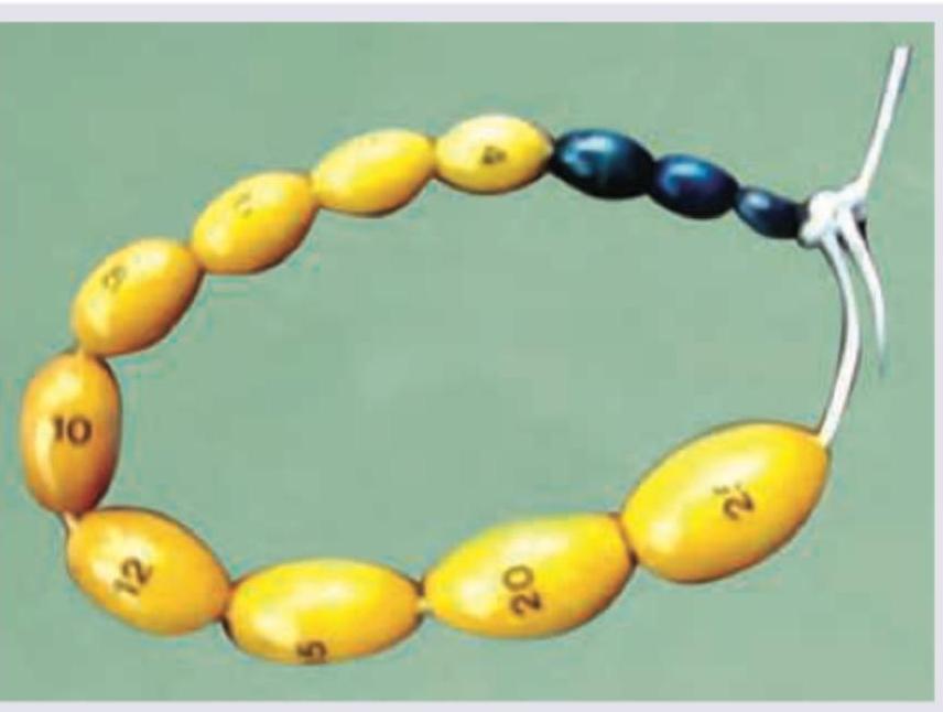
Which is true about the instrument shown?

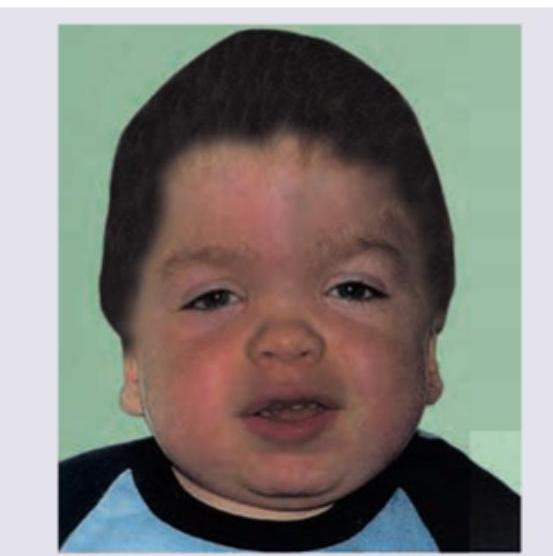
What is the diagnosis of this child with short stature and corneal opacity?

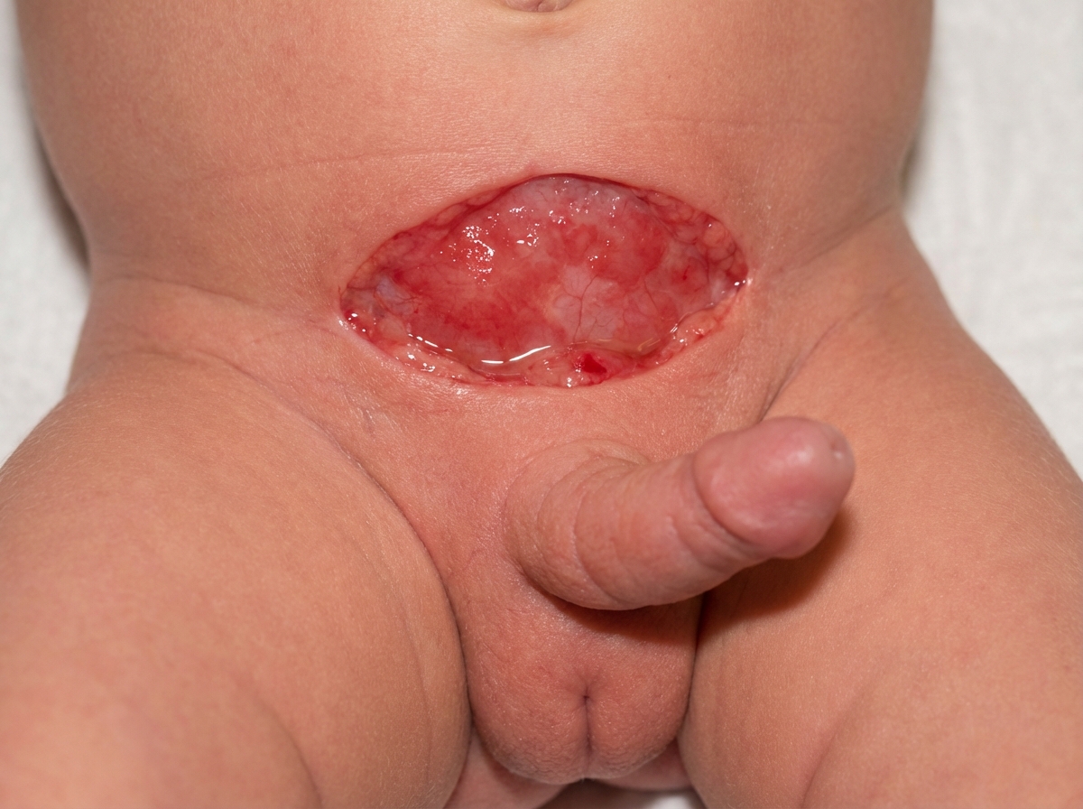
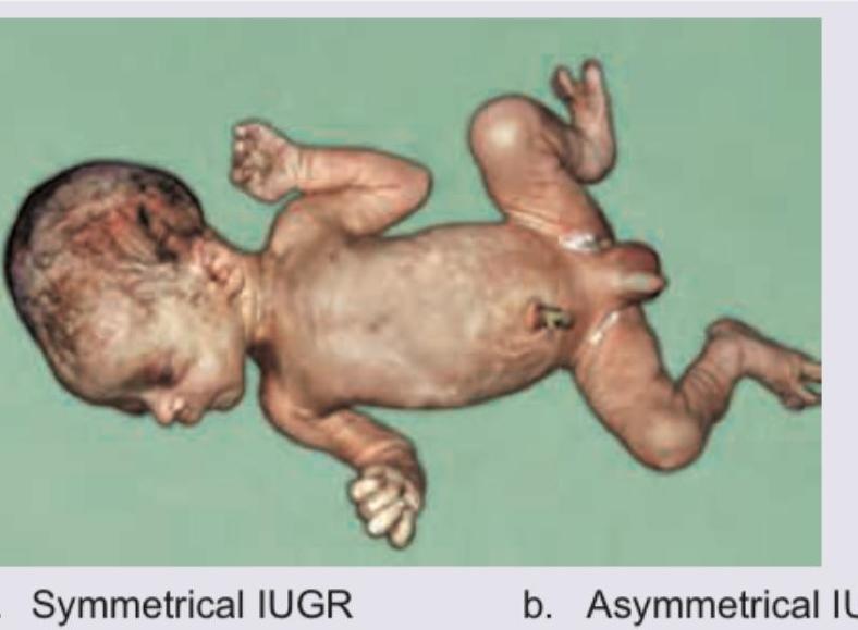
A newborn male infant presents with the findings shown in the image. The clinical diagnosis is?

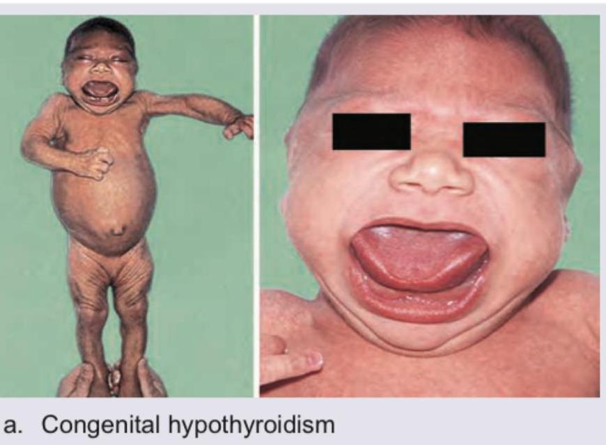
A newborn presents with macrosomia, plethoric appearance, and generalized edema. The image shows:

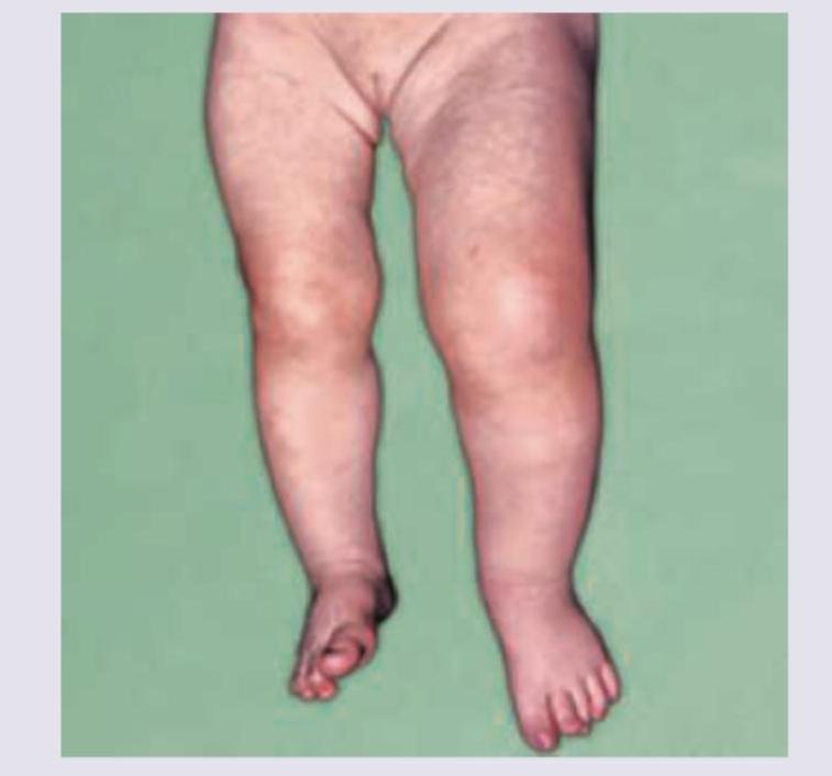
Comment on the diagnosis of the child:

All are true about this reflex except:

The reflex shown below disappears by what age?

One-year-old child with refractory epilepsy. Which is incorrect about the child in picture given below?

The image shows:

The image shows presence of:

Explanation: ***All are correct*** - This instrument, known as an **orchidometer**, is used to measure and assess **testicular volume** by comparing the patient's testicular size to the graduated ellipses. - The first three beads (1, 2, 3 ml) represent **prepubertal testicular volumes**, indicating little to no pubertal development. - Puberty is considered to have started when the testicular volume reaches **4 mL** or more, which is the established threshold for **Tanner stage 2** in male pubertal development. *Used to assess testicular volume* - The pictured instrument is an **orchidometer**, specifically designed for the clinical assessment of **testicular size** and volume. - It consists of a series of beads of increasing sizes, each labeled with a volume in milliliters, which are used to visually and tactilely compare against the patient's testicles. *First three beads are pre pubertal* - Testicular volumes of **1, 2, and 3 mL** are considered to be within the **prepubertal range**. - These volumes indicate that significant pubertal development has not yet begun. *Puberty starts at ≥4 ml* - A testicular volume of **≥4 mL** is the clinical indicator of the **onset of puberty (Tanner stage 2)** in males. - This threshold signifies the beginning of **gonadal maturation** and testicular growth associated with pubertal hormonal changes.
Explanation: ***Mucopolysaccharidosis*** - The combination of **short stature** and **corneal opacity** (corneal clouding) is pathognomonic for Mucopolysaccharidoses (MPS). - MPS are lysosomal storage disorders characterized by the accumulation of **glycosaminoglycans (GAGs)** in various tissues, leading to multi-system involvement. - Corneal clouding is particularly seen in **MPS I-H (Hurler), I-S (Scheie), IV (Morquio), and VI (Maroteaux-Lamy)**. - Other features include coarse facial features, hepatosplenomegaly, skeletal dysplasia, and developmental delay. *Cretin* - **Cretinism** (congenital hypothyroidism) presents with **short stature**, developmental delay, and coarse features. - **Corneal opacity is NOT a typical manifestation** of hypothyroidism. - Other common features include large tongue, umbilical hernia, hypotonia, and prolonged jaundice. *Peroxisomal disorders* - **Peroxisomal disorders** (e.g., Zellweger syndrome) involve neurological and developmental abnormalities with **short stature**. - While some may rarely have corneal changes, **cataracts and retinopathy** are more characteristic ocular findings. - The combination of short stature with prominent corneal opacity is not typical. *Lesch-Nyhan syndrome* - **Lesch-Nyhan syndrome** is an X-linked recessive disorder of purine metabolism causing **hyperuricemia** and severe neurological dysfunction. - It does NOT present with **short stature** or **corneal opacity**. - Characteristic features include self-mutilation behavior, dystonia, choreoathetosis, and developmental delay.
Explanation: ***Epispadias*** - The image depicts a severe form of **epispadias**, characterized by the exposed **bladder mucosa (exstrophy)** and a penile shaft that is short, dorsally curved, and has the urethral opening on the dorsal surface. - This congenital anomaly results from a failure of the abdominal wall and bladder to close properly during fetal development, often presenting with a **widened pubic symphysis**. *Hypospadias* - **Hypospadias** is a condition where the **urethral opening** is located on the **ventral** (underside) surface of the penis, not the dorsal surface as seen in the image. - While it can involve chordee (penile curvature), it typically does not present with bladder exstrophy. *Micro-penis* - **Micropenis** refers to a penis that is **abnormally small** in length, but otherwise structurally normal. - It describes the size of the penis, not an anomalous opening or exposed internal organs, and is not consistent with the image. *Mauriac syndrome* - **Mauriac syndrome** is a rare complication of poorly controlled **Type 1 diabetes** in children, characterized by growth retardation, delayed puberty, hepatomegaly, and Cushingoid features. - It is an **endocrine disorder** and has no direct relevance to the anatomical abnormality shown in the image.
Explanation: ***Infant of diabetic mother*** - The image shows a **macrosomic** infant with a **plethoric appearance**, generalized **edema**, and a **"cherubic" facial appearance** with a prominent philtrum and perioral cyanosis, all characteristic features of an infant of a diabetic mother. - Maternal diabetes leads to fetal **hyperinsulinemia**, resulting in increased fat deposition, organomegaly, and altered growth patterns. *Achondroplasia* - Achondroplasia is characterized by **proximal limb shortening** (rhizomelia), a large head with a prominent forehead, and a flattened nasal bridge. - The pictured infant does not show these specific skeletal dysplasias. *Hemi-hypertrophy* - Hemi-hypertrophy involves **asymmetric growth**, where one side or part of the body is significantly larger than the other. - The infant in the image exhibits generalized, symmetrical macrosomia rather than unilateral overgrowth. *Albright hereditary osteodystrophy* - Albright hereditary osteodystrophy is a genetic disorder associated with short stature, **obesity**, brachydactyly (short digits), and typically presents with features related to **pseudohypoparathyroidism** (e.g., hypocalcemia, hyperphosphatemia). - The characteristic findings of this syndrome, such as brachydactyly, are not evident in the image.
Explanation: ***Congenital hypothyroidism*** - The image displays characteristic features of untreated congenital hypothyroidism, including a **puffy face**, **protruding tongue** (macroglossia), and **umbilical hernia** with a distended abdomen. - These infants also typically show **poor feeding**, **lethargy**, **constipation**, **prolonged jaundice**, and **hoarse cry**, due to the widespread effects of thyroid hormone deficiency on growth and metabolism. - **Newborn screening** can detect this early, and prompt thyroid hormone replacement prevents intellectual disability. *Down syndrome* - While Down syndrome can also present with some similar facial features, such as a **protruding tongue**, it is primarily characterized by distinct facial features like an **upward slant to the eyes**, **epicanthal folds**, **flat nasal bridge**, and **low-set ears**. - Other common features include a **single palmar crease**, **hypotonia**, **congenital heart defects**, and developmental delay, which help differentiate it from congenital hypothyroidism. *Beckwith-Wiedemann syndrome* - This syndrome is characterized by **overgrowth**, specifically features like **macrosomia** (large body at birth), **macroglossia** (large tongue), **omphalocele** or umbilical hernia, **hemihyperplasia**, and **ear creases/pits**. - Although **macroglossia** and **umbilical hernia** are present, children with Beckwith-Wiedemann are typically **large for gestational age** with accelerated growth, contrasting with the **growth retardation** and **developmental delay** seen in hypothyroidism. *Hurler syndrome* - Hurler syndrome (Mucopolysaccharidosis type I) presents with **coarse facial features**, **macroglossia**, **umbilical hernia**, **hepatosplenomegaly**, and **developmental delay**. - However, distinguishing features include **corneal clouding**, **skeletal dysplasia** (dysostosis multiplex), **joint stiffness**, and **progressive deterioration**, which typically manifest more prominently after 6-12 months of age. - The puffy myxedematous appearance in hypothyroidism differs from the coarse facies in Hurler syndrome.
Explanation: ***First primitive reflex to disappear*** - The **Moro reflex** typically disappears around **3-6 months** of age, making it one of the later primitive reflexes to vanish, not the first. - Other reflexes like the **rooting reflex** (disappears around 4 months) or **stepping reflex** (disappears around 2 months) may disappear earlier or around the same time. *Embracing reflex* - The Moro reflex is also known as the **embracing reflex** due to the characteristic adduction and flexion of the limbs, as if embracing, following the initial abduction. - This name accurately describes the two-part motor response observed in infants. *Bilateral absence is seen in HIE stage III* - The **Moro reflex** can be absent bilaterally in severe neurological conditions such as **Hypoxic-Ischemic Encephalopathy (HIE) stage III**, indicating significant brain damage. - Its absence is a key indicator of **neurological dysfunction** in neonates. *Unilateral absence in Erb's palsy.* - **Unilateral absence** of the Moro reflex is often a sign of a neurological or musculoskeletal injury on one side of the body, such as **Erb's palsy**, which involves damage to the brachial plexus. - It can also be seen in cases of **fractured clavicle** or **humerus** on the affected side.
Explanation: ***3 months*** - The image depicts the **rooting reflex**, where a baby turns its head and opens its mouth in response to a touch on the cheek or around the mouth, indicating readiness to feed. - This primitive reflex is crucial for feeding in newborns and typically **disappears by 3-4 months of age**. - **3 months represents the beginning of the typical age range** when this reflex starts to disappear in neurologically normal infants. *1 month* - At 1 month of age, the rooting reflex is still prominently present and active. - This is too early for the reflex to disappear; it serves an important feeding function at this age. *2 months* - While the rooting reflex may begin to diminish slightly by 2 months, it typically persists beyond this age. - This represents an earlier disappearance than the accepted standard range of 3-4 months. *4 months* - By 4 months, the rooting reflex has usually fully integrated or disappeared in most neurologically normal infants. - While this is within the acceptable range (3-4 months), **3 months is the more commonly cited age** when disappearance typically begins. - Persistence beyond 4-6 months may warrant neurological evaluation.
Explanation: ***Geographical skull*** ✓ **Correct Answer (Incorrect about the child)** - **Geographical skull** (or "map-like skull") refers to characteristic **lytic lesions of the skull** seen in **Langerhans cell histiocytosis** (histiocytosis X). - This finding is **NOT associated with Sturge-Weber syndrome**, making this the correct answer to the question. - Sturge-Weber syndrome shows **intracranial calcifications** (tramline/railroad track sign) on imaging, not lytic lesions. *Port wine stain* ✗ **Incorrect (This IS present)** - The image shows a prominent **port-wine stain (nevus flammeus)** on the child's face, a classic cutaneous feature of **Sturge-Weber syndrome**. - This capillary vascular malformation typically follows the distribution of the **trigeminal nerve** (V1/V2 branches). *Seizures* ✗ **Incorrect (This IS present)** - **Refractory epilepsy** is a cardinal feature of Sturge-Weber syndrome, resulting from underlying **leptomeningeal angiomatosis** and progressive cortical damage. - The combination of facial **port-wine stain** and **seizures** is highly suggestive of this neurocutaneous syndrome. *Buphthalmos* ✗ **Incorrect (This CAN be present)** - **Buphthalmos** ("ox eye") refers to an enlarged eyeball associated with **congenital glaucoma**. - **Glaucoma** occurs in approximately **30-70%** of Sturge-Weber patients, particularly when the port-wine stain involves the **upper eyelid** (V1 distribution), indicating potential ocular involvement.
Explanation: ***Symmetrical IUGR*** - The image displays a neonate with **proportionately small body parts**, including the head and trunk, which is characteristic of symmetrical intrauterine growth restriction. - This type of IUGR typically results from **early gestational insults** (e.g., genetic abnormalities, infections) affecting overall cell number and size. *Asymmetrical IUGR* - Asymmetrical IUGR (also known as "head-sparing" IUGR) is characterized by a **normal head size** relative to a disproportionately small body. - This condition usually occurs due to **late-onset fetal malnutrition** or placental insufficiency, where the brain is preferentially supplied with nutrients. *Proteus syndrome* - Proteus syndrome involves **overgrowth of various tissues** (skin, bone, adipose tissue), leading to asymmetrical and disorganized growth. - The image shows a uniformly small infant, not focal or multi-tissue overgrowth, ruling out Proteus syndrome. *Poland syndrome* - Poland syndrome is a rare birth defect characterized by **missing or underdeveloped muscles on one side of the body**, primarily affecting the chest and arm. - The infant in the image does not exhibit unilateral muscle deficiency or limb abnormalities typical of Poland syndrome.
Explanation: ***Hemihypertrophy*** - The image clearly demonstrates **asymmetric growth**, with one leg appearing significantly larger and longer than the other, which is characteristic of hemihypertrophy. - This condition involves the **overgrowth of one side of the body or a body part**, and is often associated with certain genetic syndromes like Beckwith-Wiedemann syndrome. *Polydactyly* - **Polydactyly** refers to the presence of **extra fingers or toes**. - The image does not show any supernumerary digits; instead, it highlights a discrepancy in limb size. *Phocomelia* - **Phocomelia** is a rare congenital malformation where the **proximal parts of limbs are absent or severely reduced**, leading to hands or feet being attached close to the trunk. - The image displays fully formed limbs, albeit with disproportionate size. *Amniotic band sequence* - **Amniotic band sequence** results from entrapment of fetal parts in fibrous amniotic bands, leading to **constriction rings, amputations, or other deformities**. - No such bands, constrictions, or amputations are visible in the image; the primary finding is asymmetric limb growth.
Normal Growth Parameters
Practice Questions
Developmental Milestones
Practice Questions
Puberty and Adolescent Development
Practice Questions
Growth Disorders
Practice Questions
Failure to Thrive
Practice Questions
Developmental Screening and Assessment
Practice Questions
Developmental Delays
Practice Questions
Growth Charts and Monitoring
Practice Questions
Short Stature
Practice Questions
Tall Stature
Practice Questions
Precocious and Delayed Puberty
Practice Questions
Psychosocial Development
Practice Questions
Get full access to all questions, explanations, and performance tracking.
Start For Free