A 6-year-old child is brought for complaints of short stature. His height is comparable with his parent's height and his bone age corresponds to his chronological age. What is the likely diagnosis?
Gonadal growth corresponds with _____.
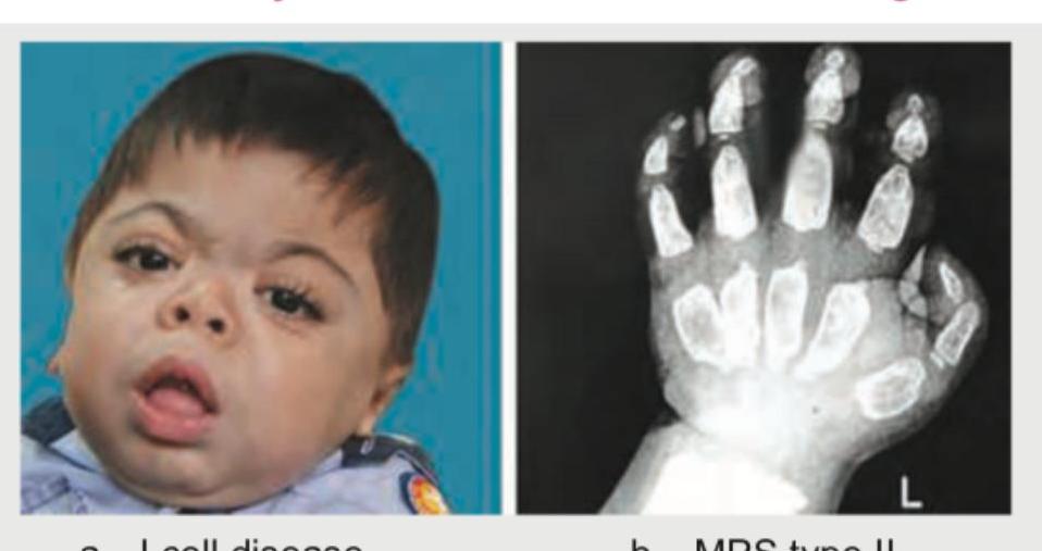
A 10-month-old child with coarse facies is referred for developmental delay. On examination, hepatosplenomegaly was noted. WBC N-acetylglucosamine-1-phosphotransferase activity was absent. The X-ray is shown below. What is the diagnosis?

A mother is concerned about the bowing of legs of her child. Which tests should be performed to label it as physiological genu varum?
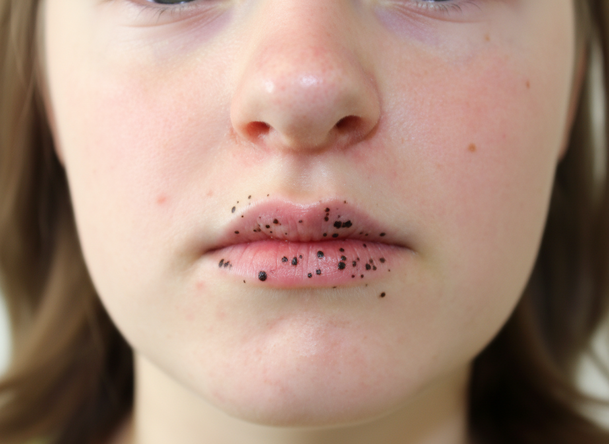
A 6-year-old child is brought with the following lesion. He can develop lesions in which organ in future?

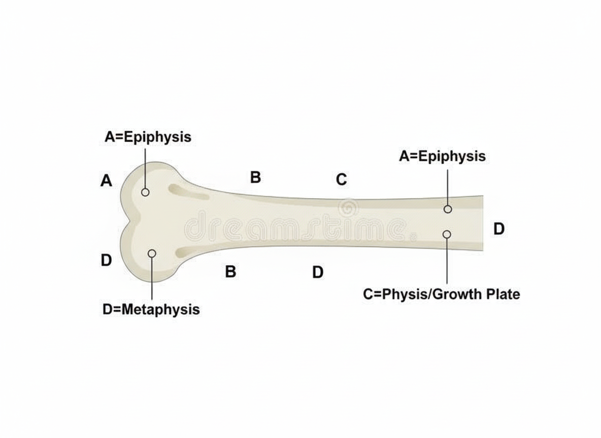
Which structure is most commonly affected in rickets?

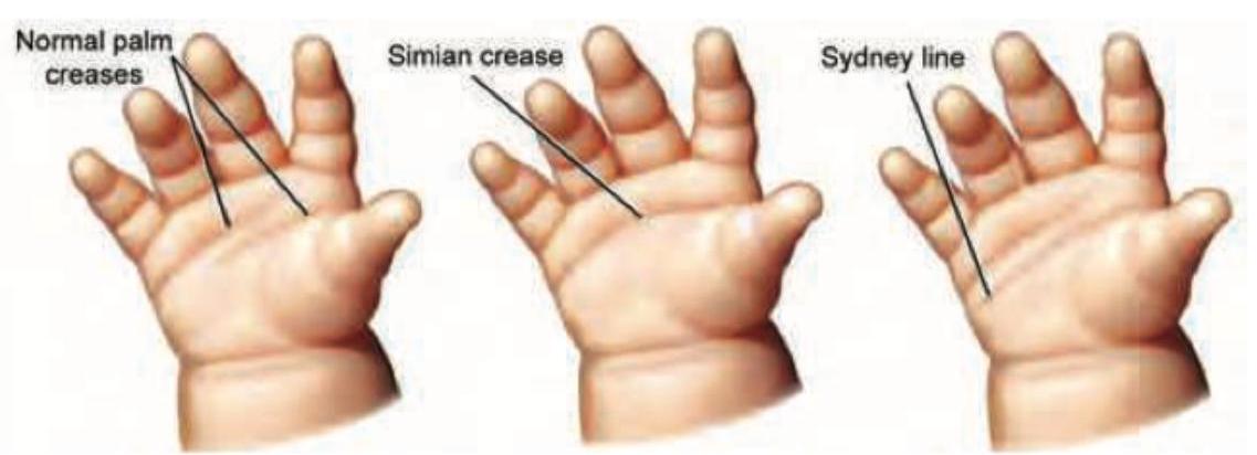
The image displays a hand with a distinct palmar crease pattern where a transverse crease extends from the radial to the ulnar border, in addition to another proximal transverse crease. What is this specific crease pattern called?

The picture depicts Rocker bottom feet. This finding is classically associated with which chromosomal abnormality?

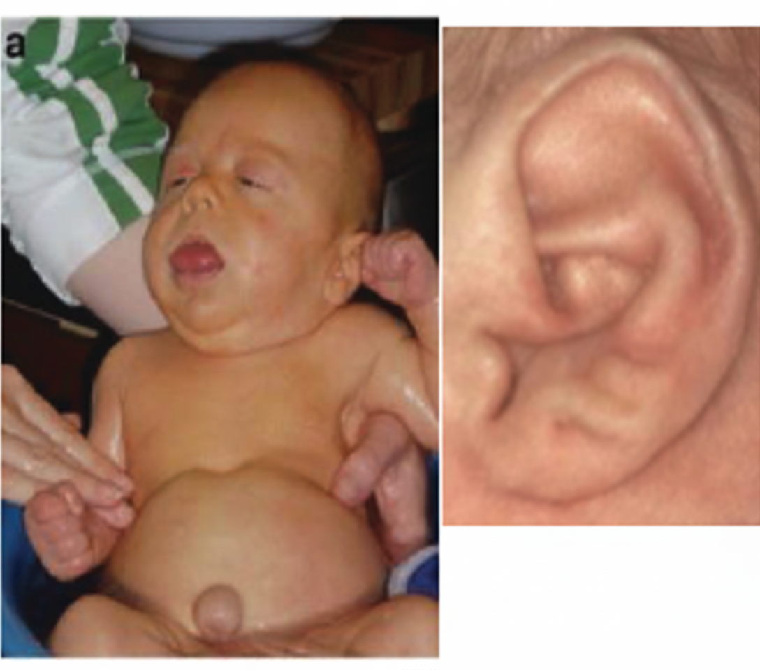
Ear lobe creases in a child are commonly associated with which disorder that also presents with macroglossia and macrosomia?

A child presents with polydactyly and obesity. Which syndrome is most likely associated with these clinical findings?

Explanation: ***Familial short stature***- The short stature is proportional, and the growth velocity is usually normal, with the child tracking below the 3rd percentile but parallel to the normal curve.- Crucially, the **bone age corresponds to the chronological age**, meaning the skeletal maturation rate is normal, and they have an adult height potential consistent with their genetic background.*Constitutional delay*- This condition is characterized by a **delayed bone age** (e.g., bone age is 4 years in a 6-year-old child), which contradicts the case findings.- These children often have delayed puberty but will eventually reach a normal adult height, simply having a late **growth spurt**.*Undernutrition*- Chronic severe undernutrition causes short stature, often presenting with features like **wasting or stunting** and usually **delayed bone maturation**.- While it is a cause of growth failure, the history of height correlating with parents and normal bone age makes primary undernutrition less likely.*Growth hormone deficiency*- GH deficiency typically results in a **severely reduced growth velocity** and often significantly **delayed bone age**.- The pattern of growth velocity and height is typically inconsistent with the mid-parental height, unlike in familial short stature.
Explanation: ***Skeletal system*** - **Gonadal growth** follows the Genital growth curve (Scammon's curves), characterized by minimal pre-pubertal growth and dramatic acceleration during **puberty** (ages 12-18). - The **skeletal system** follows the General growth curve, which similarly exhibits a significant **pubertal growth spurt** coinciding with the surge in sex hormones. - Both gonadal maturation and skeletal growth during puberty are driven by **sex steroids** (estrogen and testosterone), making their growth patterns temporally aligned and hormonally interdependent. - The timing of peak skeletal growth velocity (PHV) corresponds closely with the rapid gonadal development during **adolescence**. *Brain* - The **brain** follows the Neural growth curve, which shows rapid growth in **early childhood** (reaching ~90% of adult size by age 6 years) and then plateaus. - This pattern is **opposite** to gonadal growth, which remains relatively dormant in childhood and accelerates only during puberty. - Neural and Genital curves are the most divergent among Scammon's four growth curves. *Lymphoid growth* - **Lymphoid tissue** (thymus, lymph nodes, tonsils) follows a unique curve with rapid early growth, reaching peak size (>100% adult size) by age **10-12 years**, then involuting during puberty. - This pattern of early hypertrophy and pubertal regression is **inverse** to gonadal development. *Dental growth* - **Dental eruption** follows a predictable, age-specific schedule related to the General growth pattern and craniofacial bone development. - While influenced by overall maturation, dental development does not exhibit the characteristic hormone-dependent **pubertal acceleration** seen in gonadal growth.
Explanation: ***I cell disease*** - **I-cell disease** (Mucolipidosis II) is characterized by **coarse facial features**, developmental delay, hepatosplenomegaly, and **skeletal abnormalities** (dysostosis multiplex) seen on X-ray, which are consistent with the image. - The absence of **N-acetylglucosamine-1-phosphotransferase (GlcNAc-1-phosphotransferase)** activity leads to misrouting of lysosomal enzymes, resulting in accumulation of mucolipids within cells. *MPS type II* - **MPS type II** (Hunter syndrome) also presents with coarse facies, developmental delay, and hepatosplenomegaly, and can show skeletal abnormalities. - However, the enzymatic defect is in **iduronate sulfatase**, not GlcNAc-1-phosphotransferase, and the clinical course tends to be slightly milder than I-cell disease, particularly in early infancy. *Proteus syndrome* - **Proteus syndrome** is characterized by **overgrowth of tissues**, asymmetric growth, and various tumors, not by the specific metabolic defect or typical pattern of skeletal changes described. - It does not involve absent GlcNAc-1-phosphotransferase activity. *Larsen syndrome* - **Larsen syndrome** primarily involves **skeletal abnormalities**, such as joint dislocations (especially knees, hips, and elbows), flattened facial appearance, and clubfoot. - It does not feature the same metabolic defect (absent GlcNAc-1-phosphotransferase activity) or the prominent coarse facial features and hepatosplenomegaly seen in the patient.
Explanation: ***X-ray wrist, cover-up test, urine calcium and SAP*** - An **X-ray of the wrist** is used to assess bone age and look for metaphyseal changes suggestive of rickets, which would rule out physiological genu varum. - The **cover-up test** (specifically to evaluate for Blount disease) helps differentiate physiological bowing from pathological bowing, while **urine calcium and serum alkaline phosphatase (SAP)** are used to rule out nutritional rickets or other metabolic bone disorders. *X-ray wrist, urine hydroxyproline, urine calcium and SAP* - While X-ray wrist, urine calcium, and SAP are relevant, **urine hydroxyproline** is primarily used as a marker for bone turnover and collagen degradation and is not a primary diagnostic test for distinguishing physiological genu varum. - The inclusion of **urine hydroxyproline** makes this option less precise for the direct diagnosis of physiological genu varum compared to other tests. *X-ray wrist, urine aminoaciduria* - An **X-ray of the wrist** is appropriate, but **urine aminoaciduria** is a marker for certain metabolic disorders affecting renal reabsorption or amino acid metabolism, not directly for ruling out common causes of pathological bowing or confirming physiological genu varum. - This option lacks tests that specifically screen for metabolic bone diseases like rickets, which are crucial differentiators. *X-ray wrist, urinary calcium* - While both parameters are relevant, this option is **incomplete** as it misses important diagnostic tools. - **Serum alkaline phosphatase (SAP)** is crucial for evaluating bone turnover and rickets, and a specific clinical test like the **cover-up test** or examination for other signs of Blount disease is necessary for differentiating physiological bowing.
Explanation: ***Small intestine*** - The image shows **perioral melanotic macules**, specifically around the mouth, which are classic cutaneous manifestations of **Peutz-Jeghers syndrome**. - Peutz-Jeghers syndrome is an **autosomal dominant disorder** characterized by these mucocutaneous pigmentations and an increased risk of developing **hamartomatous polyps** in the gastrointestinal tract. - The **small intestine** is the **most commonly affected site** for polyps in Peutz-Jeghers syndrome, with **60-90% of patients** developing small bowel polyps, particularly in the **jejunum**. - These polyps can cause complications including **intussusception**, bleeding, and obstruction. - Patients also have an increased risk of **malignancies** in the small intestine and other organs (breast, ovary, pancreas, colon). *Colon (Large Intestine)* - While colonic polyps do occur in Peutz-Jeghers syndrome (**20-30% of cases**), they are **less frequent** than small intestinal polyps. - The colon is a secondary site of involvement compared to the small intestine. - Increased risk of colorectal cancer exists but the predominant polyp site remains the small bowel. *Stomach* - Gastric polyps occur in **approximately 25% of patients** with Peutz-Jeghers syndrome. - While gastric involvement can occur, it is **less common** than small intestinal involvement. - The stomach is a secondary site compared to the small intestine. *Esophagus* - Esophageal involvement with polyps in Peutz-Jeghers syndrome is **very rare**. - The typical distribution of hamartomatous polyps extends from stomach to rectum, with minimal esophageal involvement. - Esophagus is the least commonly affected GI organ in this syndrome.
Explanation: ***C (Physis/Growth Plate)*** - **Rickets** is a metabolic disorder characterized by inadequate mineralization of growing bones, most commonly affecting the **physis (growth plate)** of long bones - The growth plate is the cartilaginous area between the epiphysis and metaphysis where active bone growth occurs - Defective mineralization at this site leads to widening of the growth plate and characteristic skeletal deformities - **Reference:** O. P Ghai Textbook of Pediatrics, 8th Ed, page 113 *A (Epiphysis)* - The epiphysis is the bony section at the end of a long bone - While mineralization can be affected, it is not the **primary site** of pathology in rickets - The epiphysis and physis are anatomically distinct structures *B (Diaphysis)* - The diaphysis is the shaft of long bones - Though cortical bone can show changes in rickets, the diaphysis is not the **most commonly affected** area - The actively growing physis shows more pronounced changes *D (Metaphysis)* - The metaphysis is adjacent to the growth plate and can show radiological changes (cupping, fraying) - However, the **primary pathology** originates at the physis itself - Metaphyseal changes are secondary to physeal involvement
Explanation: ***Sydney line*** - The image illustrates a hand with a distinct palmar crease pattern where a **transverse crease extends from the radial to the ulnar border**, in addition to another proximal transverse crease. - This specific crease pattern, where the proximal transverse crease reaches the ulnar border of the hand, is known as a **Sydney line** and is often associated with certain chromosomal abnormalities like Down syndrome, though it can also occur in healthy individuals. *Simian crease* - A **simian crease**, also known as a single transverse palmar crease, involves only **one prominent crease spanning across the entire palm**, often replacing the usual two transverse creases. - The image shows a hand with **two distinct transverse creases**, one extending completely across and another shorter one, meaning it is not a simian crease. *Sandal gap* - A **sandal gap** refers to a **wide gap between the first and second toes**, which is a feature of the foot, not the hand. - This term is therefore **irrelevant** to the crease pattern observed on a hand. *Kennedy crease* - The term **Kennedy crease** is **not a recognized medical or anatomical term** for a palmar crease pattern. - This option is a **distractor** and does not describe any known crease variation.
Explanation: ***Edward syndrome*** - **Rocker-bottom feet** are a classic skeletal anomaly observed in infants with **Edward syndrome (trisomy 18)**. - Other distinctive features of Edward syndrome include **micrognathia**, prominent occiput, and **overlapping fingers**. *Down syndrome* - Characterized by a **single palmar crease**, **upslanting palpebral fissures**, and a flattened facial profile. - **Rocker-bottom feet** are not typically associated with Down syndrome. *Patau syndrome* - Associated with severe malformations including **microophthalmia**, **cleft lip/palate**, **polydactyly**, and central nervous system anomalies. - While foot anomalies can occur, **rocker-bottom feet** are less characteristic compared to Edward syndrome. *Turner syndrome* - This is a **monosomy X (45, XO)** condition primarily affecting females. - Key features include **short stature**, **webbed neck**, and **ovarian dysgenesis**, not rocker-bottom feet.
Explanation: ***Beckwith-Wiedemann syndrome*** - **Beckwith-Wiedemann syndrome** is characterized by conditions such as **macrosomia** (large body size), **macroglossia** (enlarged tongue), **visceromegaly**, and **ear lobe creases** or pits. - It is an overgrowth disorder often associated with an increased risk of certain childhood cancers like **Wilms tumor** and hepatoblastoma. *Down syndrome* - **Down syndrome** (Trisomy 21) presents with distinct facial features like a **flat nasal bridge**, **epicanthal folds**, and a single palmar crease, but **ear lobe creases** are not a primary characteristic. - While **macroglossia** can be seen, **macrosomia** is generally not a feature; instead, individuals with Down syndrome often have growth delays. *Turner syndrome* - **Turner syndrome** (XO karyotype) is characterized by features such as **short stature**, a **webbed neck**, shield chest, and **low-set ears**, but not typically ear lobe creases or macroglossia. - Affected individuals are phenotypically female and experience **gonadal dysgenesis**. *Noonan syndrome* - **Noonan syndrome** shares some features with Turner syndrome, including **short stature**, **webbed neck**, and **pectus excavatum**, but also presents with distinct cardiac defects. - While it can involve various facial dysmorphia, **ear lobe creases**, **macroglossia**, and **macrosomia** are not typically defining characteristics.
Explanation: ***Laurence-Moon-Bardet-Biedl syndrome*** - This syndrome often presents with **polydactyly**, **obesity**, **retinal degeneration**, learning disabilities, and renal abnormalities. - The combination of polydactyly and obesity in a child points strongly toward this diagnosis. *Apert syndrome* - Characterized by **craniosynostosis** (premature fusion of skull bones) and **syndactyly** (fusion of fingers or toes), not typically polydactyly. - Obesity is not a primary feature of Apert syndrome. *Trisomy 21* - Also known as **Down syndrome**, it typically presents with characteristic facial features, intellectual disability, and often congenital heart defects. - While some hand anomalies can occur, **polydactyly** and **obesity** are not typical primary diagnostic features. *Fetal hydantoin syndrome* - Caused by exposure to **phenytoin** during pregnancy, leading to features like broad nasal bridge, cleft lip/palate, developmental delay, and hypoplastic fingernails. - **Polydactyly** and **obesity** are not characteristic features of this syndrome.
Normal Growth Parameters
Practice Questions
Developmental Milestones
Practice Questions
Puberty and Adolescent Development
Practice Questions
Growth Disorders
Practice Questions
Failure to Thrive
Practice Questions
Developmental Screening and Assessment
Practice Questions
Developmental Delays
Practice Questions
Growth Charts and Monitoring
Practice Questions
Short Stature
Practice Questions
Tall Stature
Practice Questions
Precocious and Delayed Puberty
Practice Questions
Psychosocial Development
Practice Questions
Get full access to all questions, explanations, and performance tracking.
Start For Free