
Gastroenterology — MCQs

On this page
A neonate with Down syndrome presents with bilious vomiting. Which of the following is the most likely diagnosis?
In neonatal cholestasis, what percentage of direct bilirubin is typically considered elevated?
Which of the following is NOT true about neonatal hepatitis?
A 10-day-old infant presents with projectile vomiting. His mother states that the infant will actively drink his milk, but he forcefully vomits after each feeding. The infant shows signs of failure to thrive, with weight loss, dehydration, and lethargy. Physical examination reveals a firm, nontender, mobile, "olive-shaped" epigastric mass. Which of the following is the most likely diagnosis?
A 5-year-old child presents with watery diarrhea for 7 days. On examination, the child's weight is 10 kg and shows hanging skin folds with a normal skin pinch. What should be the sodium concentration in the oral rehydration solution (ORS)?
For a neonate at 48 hours of age with a history of non-passage of meconium, what is the next step in evaluation?
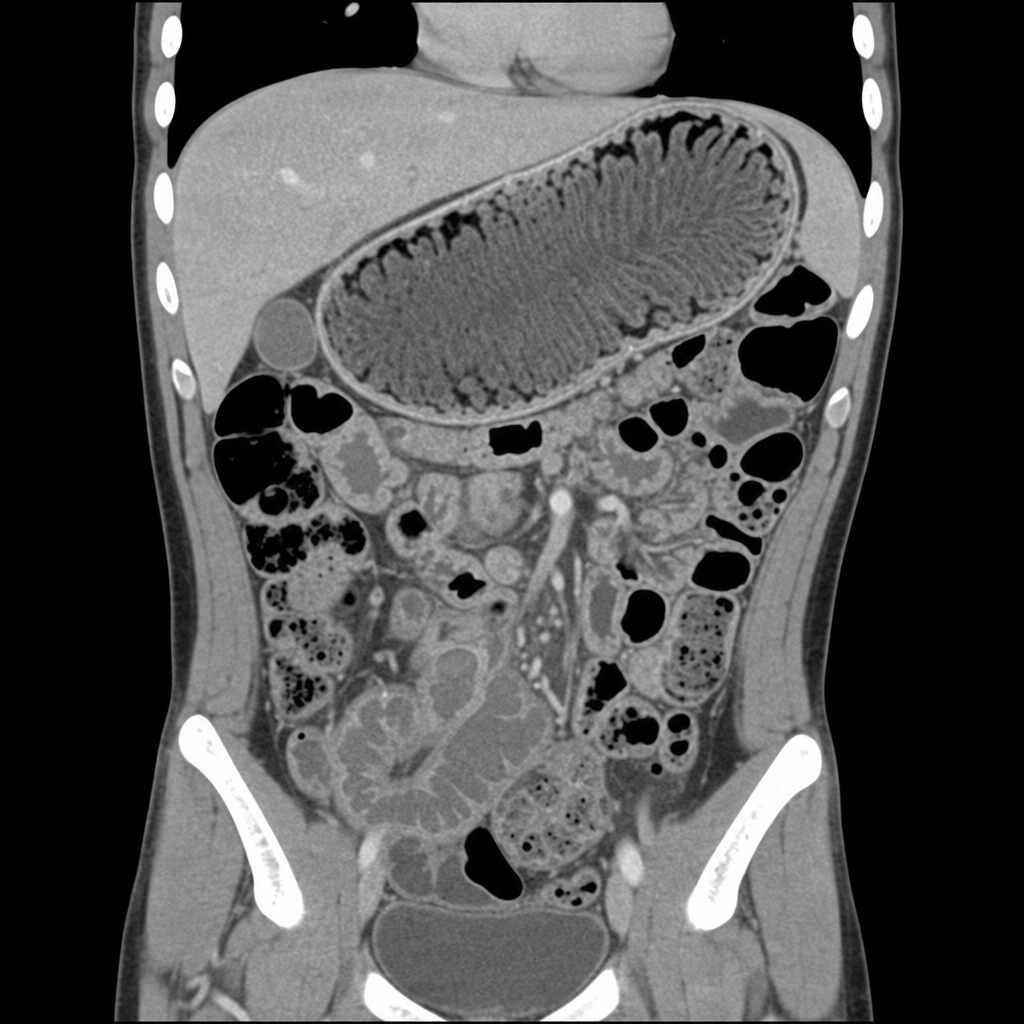
A previously healthy 5-year-old boy presented with a 4-day history of nausea, vomiting, and intermittent abdominal pain. On examination, he had mild periorbital edema. A computed tomography scan with contrast demonstrated specific findings. What is the diagnosis?
A 6-year-old boy presents with a palpable abdominal mass in the epigastrium. There is no bile in the vomitus. What is the clinical diagnosis?
In children, the presence of increased fecal fat excretion and increased fecal nitrogen levels is a feature of all the following conditions except?
What percentage of weight loss in infants constitutes severe dehydration?
Practice by Chapter
Gastroesophageal Reflux
Practice Questions
Peptic Ulcer Disease
Practice Questions
Inflammatory Bowel Disease
Practice Questions
Celiac Disease
Practice Questions
Malabsorption Syndromes
Practice Questions
Acute and Chronic Diarrhea
Practice Questions
Constipation and Encopresis
Practice Questions
Gastrointestinal Bleeding
Practice Questions
Intestinal Obstruction
Practice Questions
Liver Diseases in Children
Practice Questions
Pancreatic Disorders
Practice Questions
Pediatric Nutritional Support
Practice Questions
Want unlimited practice?
Get full access to all questions, explanations, and performance tracking.
Scan to download app