
Gastroenterology — MCQs

On this page
In a child with acute liver failure, the most important abnormal serum biochemical test that indicates poor prognosis is?
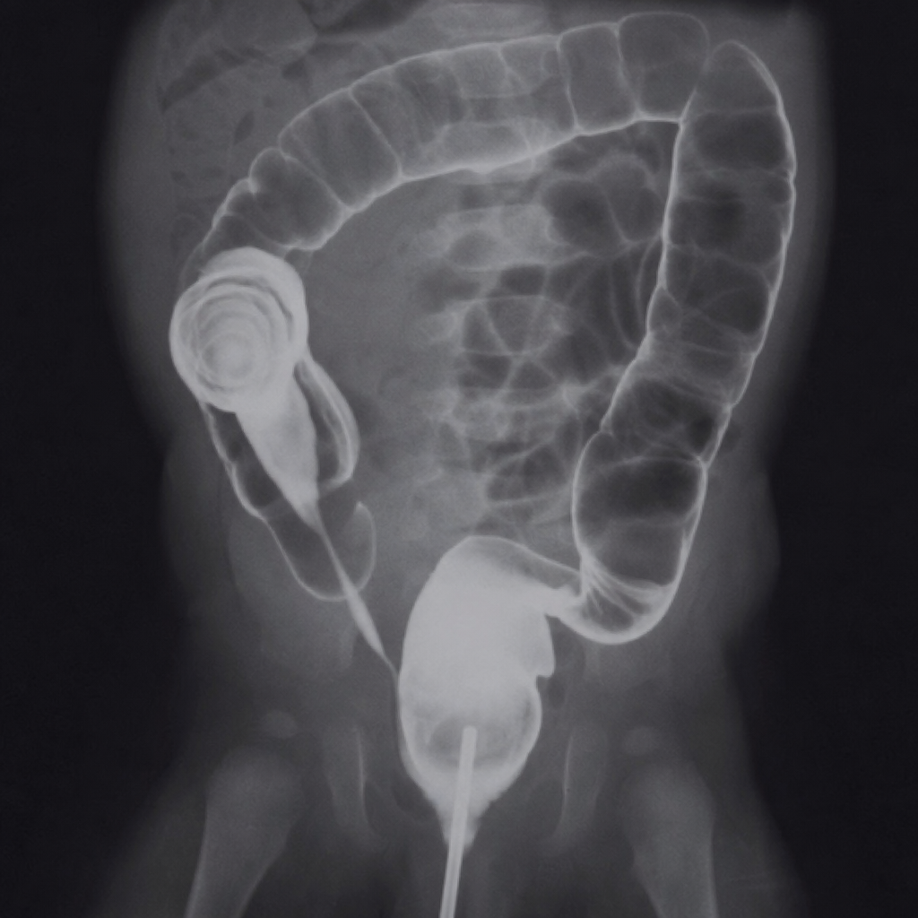
A 10 month old infant presents with acute intestinal obstruction. Contrast enema X-ray shows the intussusception, likely cause is -
A 12 year old girl has history of recurrent bulky stools and abdominal pain since 3 year of age. She has moderate pallor and her weight and height are below the 3rd percentile. Which of the following is the most appropriate investigation to make a specific diagnosis?
Which of the following is not elevated in a child presenting with jaundice, icterus, pruritus and clay-colored stools?
A previously healthy infant presents with recurrent episodes of abdominal pain. The mother says that the child has been passing altered stool after episodes of pain. There is no history of projectile vomiting or significant active bleeding per rectum. Which of the following is the most likely diagnosis -
A 10-month-old infant presents with acute intestinal obstruction. Contrast enema X-ray shows intussusception, likely cause is –
A 5-year-old child is having acute liver failure. Which one of the following criteria is not included in the King's College criteria?
In infantile hypertrophic pyloric stenosis, which metabolic disturbance is typical?
A 3-month-old with projectile vomiting and olive-shaped mass in abdomen is diagnosed with?
Most reliable indicator of some dehydration?
Practice by Chapter
Gastroesophageal Reflux
Practice Questions
Peptic Ulcer Disease
Practice Questions
Inflammatory Bowel Disease
Practice Questions
Celiac Disease
Practice Questions
Malabsorption Syndromes
Practice Questions
Acute and Chronic Diarrhea
Practice Questions
Constipation and Encopresis
Practice Questions
Gastrointestinal Bleeding
Practice Questions
Intestinal Obstruction
Practice Questions
Liver Diseases in Children
Practice Questions
Pancreatic Disorders
Practice Questions
Pediatric Nutritional Support
Practice Questions
Want unlimited practice?
Get full access to all questions, explanations, and performance tracking.
Scan to download app