
Gastroenterology — MCQs

On this page
Most common cause of significant lower GI bleeding (hematochezia) in children is
A 5 year old boy is detected to be HBsAg positive on two separate occasions during a screening program for hepatitis B. He is otherwise asymptomatic. Child was given three doses of recombinant hepatitis B vaccine at the age of 1 year. His mother was treated for chronic hepatitis B infection around the same time. The next relevant step for further investigating the child would be to –
A child is being assessed for possible intussusception; which of the following would be LEAST likely to provide valuable information?
Exocrine pancreatic insufficiency is seen in:
Which one of the following is the LEAST consistent with a diagnosis of intussusception?
In a case of hypertrophic pyloric stenosis, which of the following metabolic disturbances are found?
Organic causes of constipation in infants include all of the following EXCEPT:
Congenital pyloric stenosis causes: 1. Bilious vomiting 2. Non-bilious vomiting 3. Projectile vomiting 4. Non-projectile vomiting 5. Forceful vomiting
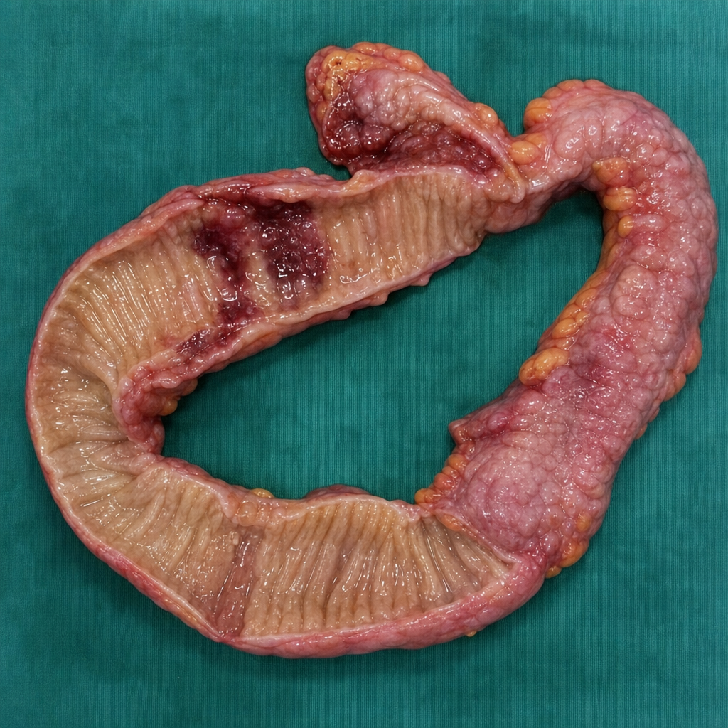
A 5-year-old boy presented with abdominal distension, fever, bilious vomiting and constipation. He also has a history of recurrent episodes of severe pain abdomen, tenesmus and blood in stools. Exploratory laparotomy was done and a poion of intestine had to be resected, which is seen below. What is the probable diagnosis?
A 7-year-old girl from Bihar presented with three episodes of massive hematemesis and melena. There is no history of jaundice. On examination, she had a large spleen, non-palpable liver, and mild ascites. Portal vein was not visualized on ultrasonography. Liver function tests were normal and endoscopy revealed esophageal varices. The most likely diagnosis is –
Practice by Chapter
Gastroesophageal Reflux
Practice Questions
Peptic Ulcer Disease
Practice Questions
Inflammatory Bowel Disease
Practice Questions
Celiac Disease
Practice Questions
Malabsorption Syndromes
Practice Questions
Acute and Chronic Diarrhea
Practice Questions
Constipation and Encopresis
Practice Questions
Gastrointestinal Bleeding
Practice Questions
Intestinal Obstruction
Practice Questions
Liver Diseases in Children
Practice Questions
Pancreatic Disorders
Practice Questions
Pediatric Nutritional Support
Practice Questions
Want unlimited practice?
Get full access to all questions, explanations, and performance tracking.
Scan to download app