A 3-month-old with projectile vomiting and olive-shaped mass in abdomen is diagnosed with?
A 24-day-old neonate presents with projectile vomiting and failure to gain weight. What is the most likely diagnosis?
Gold standard investigation for diagnosing GERD:
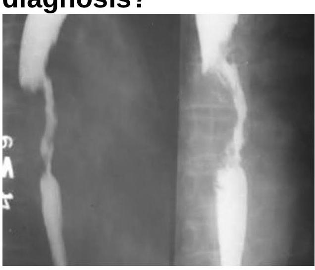
A 55-year-old male presents with a history of dysphagia with vomiting of undigested food throughout the day, weight loss, and appears emaciated and dehydrated. No mass is palpable per abdomen. After appropriate diagnostic workup reveals a benign esophageal stricture, the most appropriate definitive management is:
The Barium Swallow examination shows a filling defect in the esophagus. What is the most probable diagnosis?

A 40-year-old male with gastroesophageal reflux disease (GERD) is found to have Barrett's esophagus with low-grade dysplasia on endoscopy. What is the most appropriate initial pharmacological treatment for this condition?
A 60-year-old man presents with foul breath and regurgitates food eaten 3 days ago. What is the most likely diagnosis?
The most common complication of Zenker's diverticulum is:
False statement about Barrett esophagus is:
Adenocarcinoma of the esophagus is commonly found in patients with which of the following conditions?
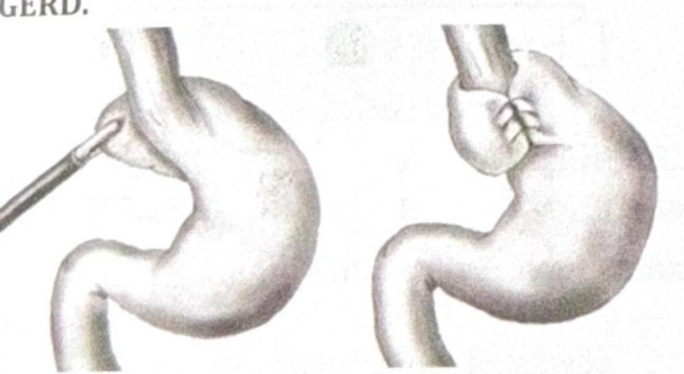
Explanation: ***Pyloric stenosis*** - The classic triad of **projectile vomiting**, a palpable **olive-shaped mass** (hypertrophied pylorus), and age of presentation (2-8 weeks, though 3 months is still possible) are highly indicative of **pyloric stenosis**. - This condition involves thickening of the **pyloric muscle**, leading to gastric outlet obstruction and non-bilious emesis. *Hirschsprung disease* - This typically presents with **constipation**, **abdominal distension**, and failure to pass meconium, rather than projectile vomiting. - It results from the absence of **ganglion cells** in the distal colon, causing functional obstruction. *GERD* - While GERD can cause vomiting in infants, it is usually not **projectile** and is rarely associated with a palpable **olive-shaped mass**. - Infants with GERD typically respond to conservative measures like thickening feeds or acid suppressants. *Duodenal atresia* - This condition presents with **bilious vomiting** (as the obstruction is distal to the ampulla of Vater) usually within the first 24-48 hours of life. - An abdominal X-ray would show a **double bubble sign**, which is not mentioned in the presentation for pyloric stenosis.
Explanation: ***Congenital Hypertrophic Pyloric Stenosis*** - The classic presentation includes **projectile, non-bilious vomiting** in a neonate around 2-8 weeks old, leading to **failure to thrive**. - An **olive-shaped mass** (hypertrophied pylorus) may be palpable in the epigastrium. *NEC* - **Necrotizing enterocolitis (NEC)** is an inflammatory disease of the intestine, primarily affecting premature infants. - Symptoms typically include **abdominal distension**, bloody stools, and lethargy, rather than projectile vomiting. *Duodenal atresia* - Presents with **bilious vomiting** within the first 24-48 hours of life due to an obstruction below the ampulla of Vater. - An X-ray would show a **"double bubble" sign**, which is not implied by the provided symptoms. *Hirschsprung's disease* - Characterized by **failure to pass meconium** within the first 24-48 hours and chronic constipation. - Vomiting, if present, is usually **bilious** and associated with abdominal distension, not projectile in nature.
Explanation: ***24 hour pH monitoring*** - This is considered the **gold standard** for diagnosing GERD because it directly measures the frequency and duration of **acid reflux events** into the esophagus. - It helps correlate symptoms with reflux episodes, providing objective evidence for the diagnosis and guiding treatment. *USG* - **Ultrasound (USG)** is primarily used for imaging abdominal organs like the **gallbladder**, liver, and kidneys, not for directly assessing esophageal acid reflux. - While it can sometimes detect complications, it cannot diagnose the presence or severity of GERD itself. *HIDA* - **HIDA scan** (hepatobiliary iminodiacetic acid scan) is used to diagnose problems of the **gallbladder** and bile ducts, such as cholecystitis or biliary obstruction. - It is not relevant for the diagnosis of gastroesophageal reflux disease. *Manometry* - **Esophageal manometry** measures the **pressure and coordination of esophageal muscle contractions**, assessing motility disorders [1]. - While it can identify related conditions like achalasia or ineffective peristalsis, it does not directly measure acid reflux and therefore is not the gold standard for GERD diagnosis.
Explanation: ***Endoscopic dilation (preferred treatment)*** - **Endoscopic dilation** directly addresses the underlying problem of the **benign esophageal stricture** by widening the narrowed esophagus, which is crucial for relieving dysphagia and improving nutritional intake. - Given the patient's severe symptoms like **weight loss**, **emaciation**, and **dehydration**, dilation allows for symptom relief and subsequent rehydration and nutritional support. *IV total parenteral nutrition* - While TPN provides nutrition, it does not resolve the **mechanical obstruction** caused by the stricture and carries risks such as infection and metabolic complications. - It's typically reserved for situations where enteral feeding is not possible or adequate after addressing the obstruction. *IV normal saline* - **IV normal saline** would help address the immediate **dehydration**, but it does not treat the underlying cause of the patient's symptoms (the esophageal stricture). - This is a supportive measure, not the primary management strategy for the stricture itself. *pH monitoring* - **pH monitoring** is used to diagnose and assess gastroesophageal reflux disease (**GERD**), which can sometimes cause strictures. - However, in a patient with a confirmed benign esophageal stricture and severe obstructive symptoms, addressing the stricture mechanically (dilation) takes precedence over diagnostic testing for reflux.
Explanation: ***Esophageal Carcinoma*** - A filling defect on a barium swallow study, especially with irregular borders and luminal narrowing, is highly suggestive of an **esophageal carcinoma**. - The image appears to show an **irregular, obstructing lesion** that displaces the barium column, characteristic of a mass. *Esophageal Ring* - An esophageal ring, such as a **Schatzki ring**, typically presents as a thin, circumferential narrowing of the distal esophagus, forming a smooth, shelf-like indentation, which is not seen here. - Esophageal rings usually cause **intermittent dysphagia** to solids but do not present as a large, irregular filling defect. *Esophageal Tear* - An esophageal tear (e.g., **Mallory-Weiss tear**) is a mucosal laceration that would present with **hematemesis** and would typically appear as a linear defect or streak on a barium swallow if visible, not a filling defect. - A tear is not usually associated with a persistent mass effect or irregular luminal obstruction seen in the image. *Achalasia Cardia* - **Achalasia** is characterized by the failure of the lower esophageal sphincter to relax and **absent peristalsis** in the esophageal body, leading to a classic "bird's beak" or "rat tail" appearance on barium swallow due to distal narrowing and proximal dilation. - While it causes luminal narrowing, it does not typically present as an irregular filling defect within the lumen, but rather as a smooth tapering of the distal esophagus.
Explanation: ***PPI*** - In patients with **GERD** and **low-grade dysplasia**, high-dose **proton pump inhibitors (PPIs)** are the initial treatment of choice to suppress acid reflux. - Continuous acid suppression can help in the regression of dysplasia and prevent its progression to higher grades. *Fundoplication* - **Fundoplication** is a surgical procedure to treat severe GERD, but it is not the primary initial treatment for low-grade dysplasia. - It might be considered if medical therapy with PPIs fails or if there are significant anatomical defects. *Esophageal resection* - **Esophageal resection** is a major surgical procedure typically reserved for **high-grade dysplasia** or **esophageal adenocarcinoma**. - It is an overly aggressive and unnecessary intervention for initial management of low-grade dysplasia. *Diet modification* - **Diet modification** is an important adjunctive therapy for GERD symptoms and overall gastric health. - However, it is generally insufficient as a standalone initial treatment for documented **low-grade dysplasia** without concurrent pharmacotherapy.
Explanation: ***Zenker's Diverticulum*** - This condition presents with a classic triad of **dysphagia**, **regurgitation of undigested food**, and **foul breath (halitosis)** due to food retention in the diverticulum. - The regurgitation of food eaten several days ago is highly characteristic, indicating significant pooling and decomposition within the **pharyngeal pouch**. *Achalasia cardia* - Characterized by **dysphagia for both solids and liquids** and regurgitation, but the regurgitated food is typically fresh or only recently ingested, not from several days prior. - The primary pathology is the **failure of the lower esophageal sphincter (LES) to relax** and loss of peristalsis in the esophageal body. *Carcinoma esophagus* - Often presents with **progressive dysphagia** (first for solids, then for liquids) and significant **weight loss**. - While regurgitation can occur, it's usually of recently ingested food and rarely associated with the severe halitosis from long-standing food decomposition seen in Zenker's. *Loss of tone of upper esophageal sphincter* - This condition would more likely lead to **regurgitation of stomach contents** into the pharynx, rather than the retention of food in a pouch. - It could contribute to **reflux symptoms** but does not explain the formation of a diverticulum or the prolonged food retention leading to foul breath.
Explanation: ***Aspiration pneumonia*** - **Aspiration pneumonia** is the **most common complication** of Zenker's diverticulum, occurring due to chronic regurgitation of food and secretions that accumulate in the diverticulum. - Patients frequently experience **nocturnal regurgitation** of undigested food, which is then *aspirated* into the airways, leading to recurrent pulmonary infections. - This is the primary reason for surgical intervention in symptomatic patients with Zenker's diverticulum. *Lung abscess* - **Lung abscess** is a more *severe* but **less common** complication that can develop as a consequence of chronic, recurrent aspiration pneumonia. - It represents a localized, necrotizing infection and is a **progression** from untreated or recurrent aspiration, rather than the initial or most frequent complication. *Dysphonia* - While **dysphonia** (hoarseness) can occur due to irritation from regurgitated contents or compression of the recurrent laryngeal nerve, it is **uncommon** as a complication. - Dysphonia is more typically associated with **GERD** or direct laryngeal pathology. *Perforation* - **Perforation** of Zenker's diverticulum is a **rare** complication that may occur spontaneously, due to impacted food, or iatrogenically during endoscopic procedures. - While serious, it is far less common than pulmonary complications from chronic aspiration.
Explanation: ***Columnar to squamous metaplasia*** - Barrett esophagus is characterized by the replacement of the normal **squamous epithelium** of the distal esophagus with **columnar epithelium** [1]. - Therefore, the statement "Columnar to squamous metaplasia" is incorrect as it describes the opposite process, making it the false statement. *Chronic GERD is the predisposing factor* - **Chronic gastroesophageal reflux disease (GERD)** causes repeated exposure of the esophageal lining to stomach acid, leading to cellular damage [1][2]. - This chronic irritation is the primary risk factor for the development of Barrett esophagus [1]. *May lead to malignancy after few years* - Barrett esophagus is a significant risk factor for the development of **esophageal adenocarcinoma** [1][3]. - The metaplastic columnar epithelium can undergo further dysplastic changes, which can progress to invasive cancer over time [2]. *Goblet cells seen on histology* - The distinctive histological feature of Barrett esophagus is the presence of **intestinal metaplasia**, which includes the identification of **goblet cells** within the columnar epithelium [1]. - These goblet cells are a key diagnostic marker for Barrett esophagus [1]. **References:** [1] Cross SS. Underwood's Pathology: A Clinical Approach. 6th ed. Common Clinical Problems From Alimentary System Disease, pp. 348-349. [2] Kumar V, Abbas AK, et al.. Robbins and Cotran Pathologic Basis of Disease. 9th ed. The Gastrointestinal Tract, pp. 764-765. [3] Kumar V, Abbas AK, et al.. Robbins and Cotran Pathologic Basis of Disease. 9th ed. The Gastrointestinal Tract, pp. 765-766.
Explanation: ***Barrett's esophagus*** - **Barrett's esophagus** is a **precancerous condition** where the stratified squamous epithelium lining the distal esophagus is replaced by metaplastic columnar epithelium [1] [2]. This metaplasia is a direct risk factor for developing **esophageal adenocarcinoma** [1] [2]. - The chronic inflammation and cellular changes associated with **gastroesophageal reflux disease (GERD)** predispose individuals to Barrett's esophagus, which then increases the risk of malignant transformation [1] [2]. *Achalasia cardia* - **Achalasia** is a disorder characterized by the inability of the lower esophageal sphincter to relax and a lack of peristalsis in the esophageal body. While it increases the risk of **squamous cell carcinoma**, it is not primarily associated with adenocarcinoma. - The exact mechanism for increased cancer risk in achalasia is thought to be related to chronic inflammation and stasis of food, which can lead to squamous cell dysplasia. *Plummer-Vinson syndrome* - **Plummer-Vinson syndrome** is a rare condition characterized by iron deficiency anemia, dysphagia (due to esophageal webs), and atrophic glossitis. It is a risk factor for **squamous cell carcinoma** of the esophagus, pharynx, and oral cavity, but not adenocarcinoma. - The esophageal webs are typically located in the proximal or mid-esophagus, and chronic irritation from dysphagia may contribute to squamous epithelial changes. *Chronic smoking* - **Chronic smoking** is a major risk factor for various cancers, including esophageal cancer. However, it is more strongly associated with **squamous cell carcinoma** of the esophagus, particularly in the upper and middle thirds. - While smoking can indirectly contribute to GERD and thus potentially Barrett's esophagus, its primary association with esophageal cancer is with the squamous cell type rather than adenocarcinoma. **References:** [1] Kumar V, Abbas AK, et al.. Robbins and Cotran Pathologic Basis of Disease. 9th ed. The Gastrointestinal Tract, pp. 764-766. [2] Cross SS. Underwood's Pathology: A Clinical Approach. 6th ed. Common Clinical Problems From Alimentary System Disease, pp. 348-349.
Get full access to all questions, explanations, and performance tracking.
Start For Free