
Cardiology — MCQs

On this page
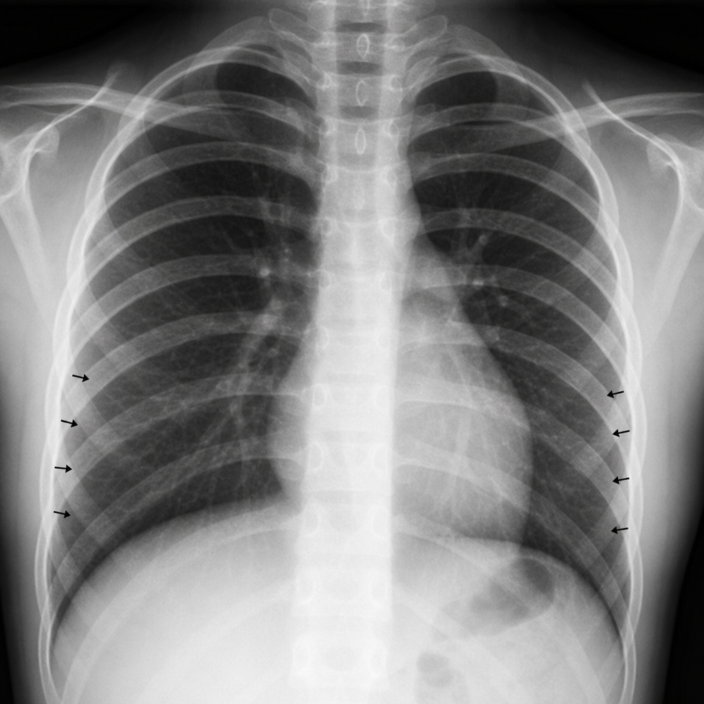
A 41/2- year-old girl always had to wear warm socks even is summer season. On physical examination, it was noticed that she had high blood pressure and her femoral pulse was weak as compared to radial and carotid pulse. a chest radiograph showed remarkable notching of ribs along with their lower borders. This was due to -
One month old baby is referred for failure to thrive. On examination there are features of congestive cardiac failure. Femoral pulses are feeble compared to brachial pulses. The likely diagnosis is
A neonate is diagnosed with pentalogy of Fallot. She may have the following lesions:
What is the most common congenital heart defect in newborns with Down syndrome?
Which among the following is a sure sign of heart failure in an infant with congenital heart disease?
A 2-week-old girl is found to have a harsh murmur along the left sternal border. The parents report that the baby gets "bluish" when she cries or drinks from her bottle. Echocardiogram reveals a congenital heart defect associated with pulmonary stenosis, ventricular septal defect, dextroposition of the aorta, and right ventricular hypertrophy. What is the appropriate diagnosis?
Tetralogy of Fallot's includes all except -
True about a 1-year-old child with PDA is –
Child with PDA will NOT have:
A patient presents with cyanosis and pulmonary complications. ECG shows left axis deviation. The most likely diagnosis is:
Practice by Chapter
Congenital Heart Diseases: Cyanotic
Practice Questions
Congenital Heart Diseases: Acyanotic
Practice Questions
Rheumatic Heart Disease
Practice Questions
Kawasaki Disease
Practice Questions
Infective Endocarditis
Practice Questions
Myocarditis and Cardiomyopathies
Practice Questions
Arrhythmias in Children
Practice Questions
Heart Failure in Children
Practice Questions
Pulmonary Hypertension
Practice Questions
Systemic Hypertension
Practice Questions
Dyslipidemia in Children
Practice Questions
Cardiac Evaluation and Diagnostics
Practice Questions
Want unlimited practice?
Get full access to all questions, explanations, and performance tracking.
Scan to download app