
Cardiology — MCQs

On this page
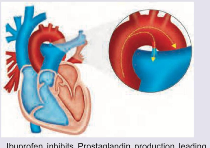
A 6-week-old term baby presents with poor feeding. All are true about the condition shown except:
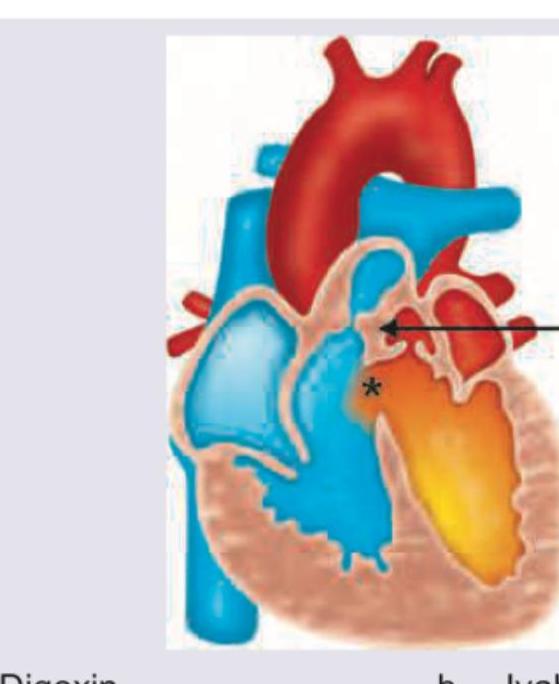
A neonate presents with a congenital heart disease as shown below. Which drug should be started immediately? (Recent NEET Pattern 2016-17)
Which congenital heart disease is associated with the defect shown?
A 1 day old neonate presents with central cyanosis. CXR and ECG are performed (shown in the image). Which diagnosis is most consistent with these findings?
Choose the TRUE statement regarding the disease depicted in the images below:
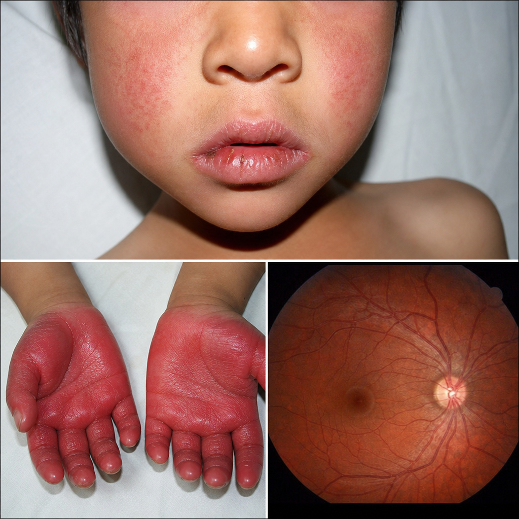
Child presents with strawberry tongue, fever for 5 days, cracked lips, Periungual peeling of the skin and bulbar congestion. Which of the following cardiac lesions will be seen in this child?
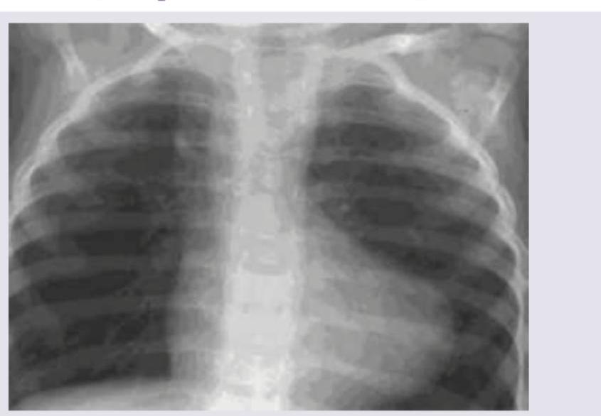
A 1-week-old neonate presents with anoxic spells and single S2. CXR shows all except:
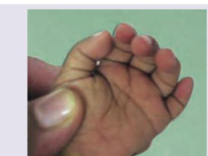
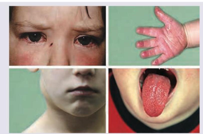
A 3-year-old child presents with the clinical features shown in the image. What is the most likely diagnosis?
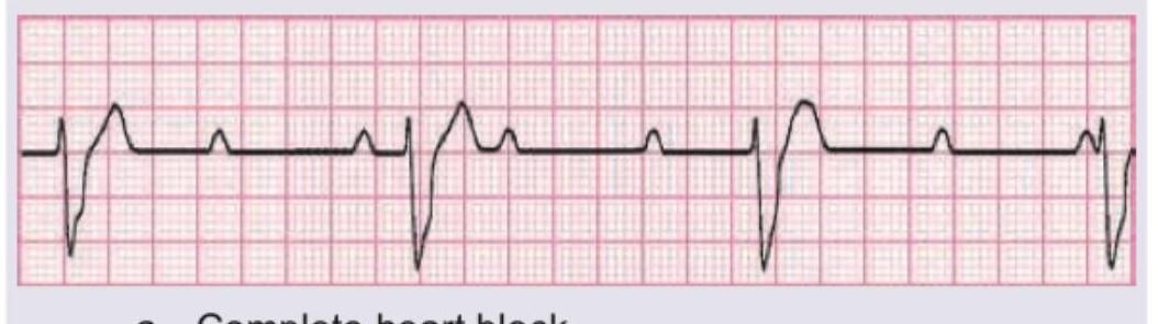
A 1-year-old child with CHD is on heart failure treatment. The ECG shows all except:
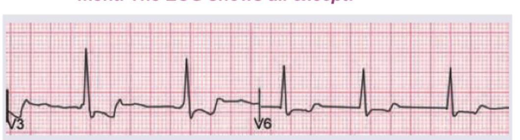
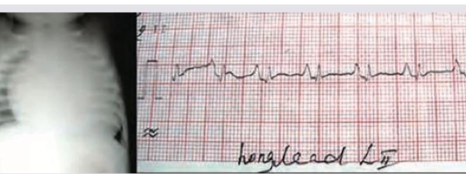
The given ECG of a neonate born to a mother with SLE shows?
Practice by Chapter
Congenital Heart Diseases: Cyanotic
Practice Questions
Congenital Heart Diseases: Acyanotic
Practice Questions
Rheumatic Heart Disease
Practice Questions
Kawasaki Disease
Practice Questions
Infective Endocarditis
Practice Questions
Myocarditis and Cardiomyopathies
Practice Questions
Arrhythmias in Children
Practice Questions
Heart Failure in Children
Practice Questions
Pulmonary Hypertension
Practice Questions
Systemic Hypertension
Practice Questions
Dyslipidemia in Children
Practice Questions
Cardiac Evaluation and Diagnostics
Practice Questions
Want unlimited practice?
Get full access to all questions, explanations, and performance tracking.
Scan to download app