Which of the following is NOT a large vessel vasculitis?
Which of the following is false about polyarteritis nodosa
Most characteristic finding in granulomatosis with polyangiitis?
A 70-year-old woman with a history of hypertension presents with a pulsatile abdominal mass. A CT scan shows an aortic aneurysm. Which histologic finding is most likely to be present in this patient?
Which of the following is not included in Virchow's triad for thrombus formation?
A red soft to firm swelling on the sternum shows proliferation of endothelial cells forming vascular channels on biopsy. What is the most likely diagnosis?
Onion skin spleen is seen in:
Which of the following is not a large vessel vasculitis?
Which of the following factors play a major role in the initiation of thrombus formation?
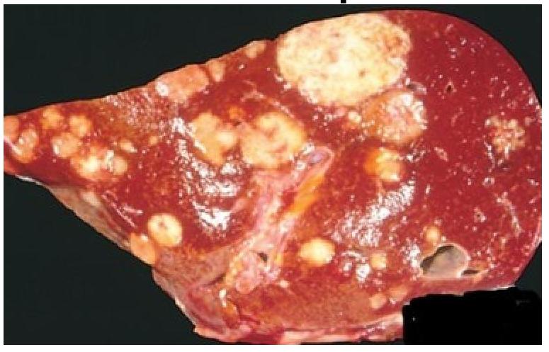
A woman shows symptoms of massive pulmonary thromboembolism. Based on the gross appearance of the liver autopsy, which of the following statements best characterizes the patient’s condition?

Explanation: ***PAN*** - **Polyarteritis Nodosa (PAN)** is a **medium-sized vessel vasculitis** that causes necrotizing inflammation of the muscular arteries [4]. - It specifically spares arterioles, capillaries, and venules, distinguishing it from large vessel vasculitides [4]. - PAN typically presents with **systemic symptoms**, **renal involvement**, **peripheral neuropathy**, and **skin manifestations** [1]. *Giant cell arteritis* - **Giant cell arteritis (GCA)**, or temporal arteritis, is a **large vessel vasculitis** affecting the aorta and its major branches, particularly the temporal artery [2]. - It commonly presents in individuals over 50 years old with symptoms like **headache**, **jaw claudication**, and **vision loss** [3]. *Aortitis* - **Aortitis** refers to inflammation of the aorta, which is the body's largest artery, making it by definition a **large vessel vasculitis**. - It can be seen in conditions like **Takayasu arteritis** [3] or **syphilis**, affecting the vessel wall. *Takayasu disease* - **Takayasu arteritis** is a chronic inflammatory condition primarily affecting the **aorta** and its main branches, classifying it as a **large vessel vasculitis** [3]. - It often affects young women and can lead to **stenosis** or **aneurysm formation** in the affected vessels [3]. **References:** [1] Cross SS. Underwood's Pathology: A Clinical Approach. 6th ed. Common Clinical Problems From Osteoarticular And Connective Tissue Disease, pp. 687-688. [2] Kumar V, Abbas AK, et al.. Robbins and Cotran Pathologic Basis of Disease. 9th ed. Diseases of Infancy and Childhood, pp. 516-517. [3] Cross SS. Underwood's Pathology: A Clinical Approach. 6th ed. Common Clinical Problems From Osteoarticular And Connective Tissue Disease, pp. 688-689. [4] Kumar V, Abbas AK, et al.. Robbins and Cotran Pathologic Basis of Disease. 9th ed. Diseases of Infancy and Childhood, pp. 517-518.
Explanation: ***Bronchial arteries are most common arteries involved*** - This statement is **FALSE** because **polyarteritis nodosa (PAN)** primarily affects **medium-sized muscular arteries**, and the **renal arteries** are the most commonly involved. - While bronchial arteries can be affected, they are not the most common target and involvement of the pulmonary circulation is rare. - PAN characteristically spares the pulmonary vasculature, which helps differentiate it from other systemic vasculitides. *ANCA negative* - **Polyarteritis nodosa (PAN)** is characterized as an **ANCA-negative vasculitis** [1]. - This feature helps differentiate it from other small-to-medium vessel vasculitides that are often ANCA-positive (e.g., GPA, MPA, EGPA) [1]. *Granulomas not seen* - **Granulomas** are a hallmark of certain vasculitides like **Granulomatosis with Polyangiitis (GPA)** but are characteristically **absent in PAN** [2]. - The inflammatory infiltrate in PAN typically consists of neutrophils, eosinophils, and lymphocytes without granuloma formation [2]. *Renal arteries are most commonly involved* - The **renal arteries** are indeed the **most frequently affected** vessels in PAN, leading to **renal ischemia**, hypertension, and potential renal failure. - Involvement of the kidneys is a major cause of morbidity and mortality in PAN. *Associated with Hepatitis B* - This statement is **TRUE** as a significant proportion of **PAN cases (20-30%)** are strongly associated with **Hepatitis B virus (HBV) infection** [1]. - **HBV-associated PAN** is thought to arise from the deposition of immune complexes containing viral antigens in vessel walls [1]. **References:** [1] Cross SS. Underwood's Pathology: A Clinical Approach. 6th ed. Common Clinical Problems From Osteoarticular And Connective Tissue Disease, pp. 687-688. [2] Kumar V, Abbas AK, et al.. Robbins and Cotran Pathologic Basis of Disease. 9th ed. Diseases of Infancy and Childhood, pp. 517-518.
Explanation: ***Geographic necrosis*** - This describes the **characteristic pattern of necrosis** seen within the granulomatous inflammation in granulomatosis with polyangiitis (GPA) [3]. - The necrosis is **basophilic, serpiginous, and irregularly shaped** (geographic pattern), surrounded by palisading histiocytes and multinucleated giant cells. - This is seen in the classic **necrotizing granulomatous inflammation** that defines GPA, particularly in the **respiratory tract** (nasal, sinus, lung) and occasionally kidneys [3]. - Geographic necrosis is the most specific histological pattern among these options for identifying GPA. *Fibrinoid necrosis* - This occurs in the walls of **small and medium-sized vessels** as part of the necrotizing vasculitis in GPA. - While important in GPA pathogenesis, fibrinoid necrosis is **not specific** to GPA—it occurs in many forms of vasculitis (polyarteritis nodosa, microscopic polyangiitis, etc.) [1], [2]. - The geographic necrosis within granulomas is more diagnostically characteristic. *Caseous necrosis* - This type of necrosis is classically associated with **tuberculosis** and some fungal infections. - It has a "cheese-like" appearance, which is distinct from the basophilic geographic pattern seen in GPA. *Coagulative necrosis* - This is a common form of necrosis associated with **ischemia** (e.g., myocardial infarction). - It preserves the architectural outline of tissue, which is not characteristic of GPA. **References:** [1] Kumar V, Abbas AK, et al.. Robbins and Cotran Pathologic Basis of Disease. 9th ed. Diseases of Infancy and Childhood, pp. 518-519. [2] Kumar V, Abbas AK, et al.. Robbins and Cotran Pathologic Basis of Disease. 9th ed. Diseases of Infancy and Childhood, pp. 517-518. [3] Kumar V, Abbas AK, et al.. Robbins and Cotran Pathologic Basis of Disease. 9th ed. Diseases of Infancy and Childhood, pp. 519-520.
Explanation: ***Medial necrosis*** - Aortic aneurysms are often associated with **medial necrosis**, a degeneration of the medial layer of the aortic wall [1]. - This finding is especially common in **syndromes** like Marfan syndrome and is critical in the pathophysiology of aneurysms. *Foamy histiocytes* - Typically associated with **atheromatous plaques** and lipid-laden lesions, not directly related to **aortic aneurysms**. - While foam cells can be seen in atherosclerosis, they do not indicate the structural changes in the aortic wall characteristic of aneurysms. *Hyaline arteriolosclerosis* - Characterized by **hyaline changes** in small arterioles, mainly related to systemic hypertension and renal complications. - This histological finding is not specific or commonly linked to **aortic aneurysms**, making it irrelevant in this context. *Lipid-laden macrophages* - Found predominantly in areas of **atherosclerosis**, indicating lipid accumulation; however, this is not a primary feature of **aortic aneurysms**. - Their presence does indicate atherosclerotic disease but does not reflect the pathological changes in an aortic aneurysm. **References:** [1] Cross SS. Underwood's Pathology: A Clinical Approach. 6th ed. Common Clinical Problems From Cardiovascular Disease, pp. 267-268.
Explanation: ***Blood hypocoagulability*** - **Hypocoagulability** refers to a reduced ability of blood to clot, which would actually decrease the risk of thrombus formation. - Virchow's triad describes factors that *promote* clot formation, so a state that inhibits clotting is not part of it. *Endothelial injury* - **Endothelial injury** is a key component of Virchow's triad, as damage to the vessel wall exposes subendothelial collagen and tissue factor, initiating the coagulation cascade [2]. - This damage can be caused by physical trauma, hypertension, or inflammation. *Stasis of blood flow* - **Stasis of blood flow**, or turbulent flow, is another crucial element, as it prevents the dilution of activated clotting factors and hinders the influx of thrombin inhibitors [3]. - This allows platelets and clotting factors to accumulate and interact more readily [2]. *Blood hypercoagulability* - **Blood hypercoagulability** (also known as thrombophilia) is the third component of Virchow's triad, representing an increased tendency of the blood to clot [1]. - This can be due to genetic factors (e.g., Factor V Leiden mutation) or acquired conditions (e.g., malignancy, oral contraceptive use) [1]. **References:** [1] Kumar V, Abbas AK, et al.. Robbins and Cotran Pathologic Basis of Disease. 9th ed. Hemodynamic Disorders, Thromboembolic Disease, and Shock, pp. 133-134. [2] Kumar V, Abbas AK, et al.. Robbins and Cotran Pathologic Basis of Disease. 9th ed. Hemodynamic Disorders, Thromboembolic Disease, and Shock, pp. 132-133. [3] Cross SS. Underwood's Pathology: A Clinical Approach. 6th ed. (Basic Pathology) introduces the student to key general principles of pathology, both as a medical science and as a clinical activity with a vital role in patient care. Part 2 (Disease Mechanisms) provides fundamental knowledge about the cellular and molecular processes involved in diseases, providing the rationale for their treatment. Part 3 (Systematic Pathology) deals in detail with specific diseases, with emphasis on the clinically important aspects., pp. 142-143.
Explanation: ***Hemangioma*** - The soft to firm swelling and histological findings indicative of vascular spaces are characteristic of a **hemangioma**, often presenting as a benign vascular tumor [1][2]. - Commonly found in the **sternum**, it may show red or bluish discoloration and typically undergoes a **biopsy** revealing endothelial cell proliferation [2]. *Osteochondroma* - This is a **benign bone tumor** that arises from the growth plate but typically presents as a hard, bony mass rather than a soft swelling. - Histologically, it shows **cartilaginous cap** and is associated with bone rather than vascular structures. *Osteoid osteoma* - Usually presents as a **painful, small bone lesion** often found in the long bones, generally not in the sternum and characterized by a **nidus** of osteoid. - The biopsy would show a **central nidus** with osteoid and woven bone, not consistent with soft swellings. *Paget disease* - This chronic bone disorder involves excessive bone remodeling, leading to enlarged and deformed bones rather than a discrete swelling. - Histologically, it would show **disorganized bone** formation and is not associated with soft or firm masses like hemangiomas. **References:** [1] Kumar V, Abbas AK, et al.. Robbins and Cotran Pathologic Basis of Disease. 9th ed. Diseases of Infancy and Childhood, pp. 481-482. [2] Kumar V, Abbas AK, et al.. Robbins and Cotran Pathologic Basis of Disease. 9th ed. Diseases of Infancy and Childhood, pp. 523-524.
Explanation: ***SLE*** - **Onion skin spleen** is a characteristic pathological finding in **Systemic Lupus Erythematosus (SLE)**, representing concentric perivascular fibrosis in the splenic arterioles. - This fibrosis is thought to be due to immune complex deposition and chronic inflammation around blood vessels. *ITP* - **Idiopathic Thrombocytopenic Purpura (ITP)** is characterized by **immune-mediated destruction of platelets**, leading to low platelet counts. - While the spleen is involved in platelet destruction in ITP, it does not typically show the "onion skin" appearance; its pathology is usually normal or shows only signs of increased platelet phagocytosis. *Thalassemia* - **Thalassemia** is a group of inherited blood disorders characterized by **reduced or absent synthesis of globin chains**, leading to ineffective erythropoiesis and chronic hemolysis. - The spleen in thalassemia often shows **extramedullary hematopoiesis** and **splenomegaly** due to increased destruction of abnormal red blood cells, but not onion skin fibrosis. *Scleroderma* - **Scleroderma**, or Systemic Sclerosis, is a **chronic autoimmune connective tissue disease** characterized by fibrosis of the skin and internal organs. - While scleroderma can affect various organs, **splenic involvement with onion skin fibrosis is not a typical feature**; its pathology often involves vascular changes and fibrosis in affected tissues, but not this concentric perivascular pattern in the spleen.
Explanation: **Churg-Strauss syndrome** - Also known as **Eosinophilic Granulomatosis with Polyangiitis (EGPA)**, this is primarily a **small-to-medium vessel vasculitis**. [1] - It classically presents with **asthma**, **eosinophilia**, and **granulomatous inflammation** affecting various organs. [1] *Takayasu arteritis* - This is a **large vessel vasculitis** that predominantly affects the **aorta** and its major branches. - It is often seen in **younger women** and can cause absent pulses and vascular stenoses. *Cogan syndrome* - This is a **large vessel vasculitis** characterized by **interstitial keratitis** and **vestibuloauditory dysfunction**, often impacting the aorta and great vessels. - While it can involve multiple vessel sizes, its systemic manifestations and potential for aortic involvement classify it within the spectrum of large vessel vasculitis. *Giant cell arteritis* - This is a **large vessel vasculitis** that primarily affects the **temporal artery** and other branches of the carotid artery. - It is typically seen in **older adults** and can cause headaches, jaw claudication, and vision loss. **References:** [1] Kumar V, Abbas AK, et al.. Robbins and Cotran Pathologic Basis of Disease. 9th ed. Diseases of Infancy and Childhood, pp. 519-520.
Explanation: ***Endothelial injury*** - **Endothelial injury** is a critical initiating factor in thrombus formation, leading to platelet adhesion and activation [1]. - Damage to the endothelium exposes the underlying **collagen** and **tissue factor**, which promote hemostasis and coagulation [2,3,5]. *Vasoconstriction* - While vasoconstriction can reduce blood flow and helps in minimizing blood loss, it is not a direct initiator of thrombus formation. - It primarily acts as a response to injury rather than a trigger for the **clotting mechanism**. *Coagulation cascade activation* - Activation of the coagulation cascade occurs after endothelial injury and is part of the clotting process, not the initiation [2]. - It involves various factors like fibrinogen and prothrombin but is secondary to the initial endothelial damage. *Platelets activation* - Platelet activation is a response to the exposed collagen due to endothelial injury and is not the initial trigger of thrombus formation [3,4,5]. - It occurs as a subsequent step once the endothelial injury has taken place, facilitating plug formation [4]. **References:** [1] Kumar V, Abbas AK, et al.. Robbins and Cotran Pathologic Basis of Disease. 9th ed. Hemodynamic Disorders, Thromboembolic Disease, and Shock, pp. 132-133. [2] Kumar V, Abbas AK, et al.. Robbins and Cotran Pathologic Basis of Disease. 9th ed. Hemodynamic Disorders, Thromboembolic Disease, and Shock, pp. 126-128. [3] Cross SS. Underwood's Pathology: A Clinical Approach. 6th ed. (Basic Pathology) introduces the student to key general principles of pathology, both as a medical science and as a clinical activity with a vital role in patient care. Part 2 (Disease Mechanisms) provides fundamental knowledge about the cellular and molecular processes involved in diseases, providing the rationale for their treatment. Part 3 (Systematic Pathology) deals in detail with specific diseases, with emphasis on the clinically important aspects., pp. 142-143. [4] Cross SS. Underwood's Pathology: A Clinical Approach. 6th ed. Common Clinical Problems From Blood And Bone Marrow Disease, pp. 581-582. [5] Kumar V, Abbas AK, et al.. Robbins and Cotran Pathologic Basis of Disease. 9th ed. Hemodynamic Disorders, Thromboembolic Disease, and Shock, p. 128.
Explanation: ***Colonic adenocarcinoma with metastasis*** - The presence of **massive pulmonary thromboembolism** often indicates **underlying malignancy** [2], particularly with **colonic adenocarcinoma** known to metastasize to the liver [1]. - This condition may present with **liver lesions** at autopsy, consistent with metastatic disease [1], supporting this diagnosis. *Metastasis from PE* - Pulmonary embolism (PE) itself does not typically give rise to **metastatic disease**; instead, it commonly arises from **deep vein thrombosis** (DVT) [2]. - This onfuses the cause of PE with its potential effects, lacking the **specificity** of a primary cancer origin. *Locally invaded hepatocellular carcinoma* - This option indicates a primary liver cancer impacting the liver directly, which would not cause **massive pulmonary thromboembolism** as its primary feature. - While hepatocellular carcinoma can cause some vascular complications, it does not correlate with **colonic adenocarcinoma** or metastatic patterns indicative of PE. *Angiosarcoma* - Though angiosarcoma is a **primary liver tumor**, it is rare and does not typically present with **massive pulmonary embolism** as a hallmark manifestation. - This type of cancer generally has a different clinical picture and distinct risk factors compared to **colonic adenocarcinoma**, making it an **unlikely option** in this context. **References:** [1] Kumar V, Abbas AK, et al.. Robbins and Cotran Pathologic Basis of Disease. 9th ed. Neoplasia, p. 282. [2] Kumar V, Abbas AK, et al.. Robbins and Cotran Pathologic Basis of Disease. 9th ed. The Lung, p. 705.
Atherosclerosis
Practice Questions
Hypertensive Vascular Disease
Practice Questions
Aneurysms and Dissection
Practice Questions
Vasculitis
Practice Questions
Venous Disease and Thrombosis
Practice Questions
Vascular Tumors
Practice Questions
Varicose Veins and Lymphatics
Practice Questions
Pathology of Vascular Interventions
Practice Questions
Vascular Diseases in Specific Organs
Practice Questions
Congenital Vascular Anomalies
Practice Questions
Get full access to all questions, explanations, and performance tracking.
Start For Free