What is the earliest lesion observed in atherosclerosis?
A 70-year-old man presents with abdominal pain and an abdominal mass. Angiography reveals an aneurysm of the abdominal aorta. What is the most likely cause?
A 40-year-old man presents with a 2-week history of recurrent oral ulcers, genital ulcers, intermittent arthritic pain of the knees, and abdominal pain. Physical examination reveals shallow ulcerations of the mucosa of the glans penis, as well as oral aphthous ulcers and conjunctivitis. Which of the following is the most likely diagnosis?
Magic syndrome is seen in which of the following conditions?
What is the earliest lesion observed in atherosclerosis?
Atherosclerosis initiation by fibroblast plaque is mediated by injury to?
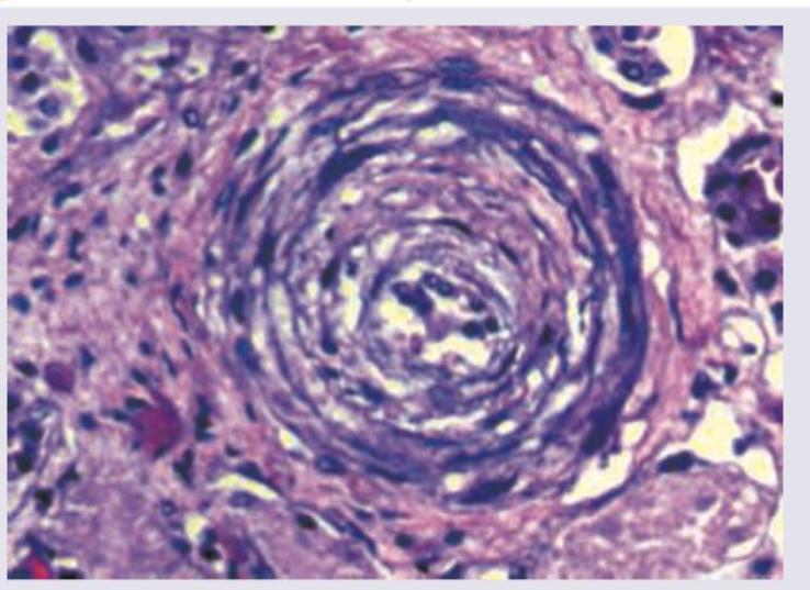
A 62-year-old man presented in the medical emergency with complaints of severe headache and dizziness. His blood pressure was recorded to be 200/146 mm Hg. Pathological slide presentation is given in the image below. What is the most likely diagnosis?

A 50-year-old patient with family history positive for premature coronary artery disease is found to have lumps on Achilles tendon. Biopsy from the lesion shows presence of:
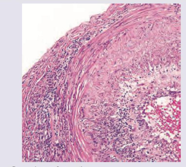
A 70-year-old retired military personnel presents with daily temporal headache and same sided blurring of vision. The Biopsy of Temporal artery was performed. All are true about the condition except:

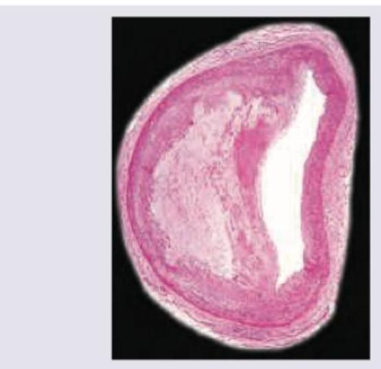
The image shows presence of:

Explanation: **Explanation:** **A. Fatty Streaks (Correct):** Fatty streaks are the **earliest visible lesion** of atherosclerosis [2]. They are composed of lipid-filled foamy macrophages (foam cells) within the tunica intima [3]. These lesions can appear as early as infancy and are present in the aortas of almost all children older than 10 years [1]. While they are precursors to more advanced lesions, not all fatty streaks inevitably progress to fibrous plaques [2]. **B. Intimal Thickening:** While some pathologists consider "pre-lesional" changes like diffuse intimal thickening to occur early, it is a non-specific response to hemodynamic stress. In the context of the standard classification of atherosclerotic lesions, fatty streaks are the first identifiable pathological stage [3]. **C. Fibrinoid Necrosis:** This is a form of vascular damage characterized by the leakage of plasma proteins (like fibrin) into the vessel wall. It is typically seen in **malignant hypertension** or **immune-mediated vasculitis** (e.g., Polyarteritis Nodosa), not as a primary stage of atherosclerosis. **D. Plaque (Atheroma):** An atherosclerotic plaque (fibrofatty plaque) is a **late-stage, mature lesion** [4]. It consists of a necrotic lipid core covered by a firm, white fibrous cap [5]. This stage represents significant progression beyond the initial fatty streak. **High-Yield NEET-PG Pearls:** * **Location:** Fatty streaks are most commonly found in the **aorta** in young individuals, whereas mature plaques are most frequent in the **lower abdominal aorta** and **coronary arteries**. * **Pathogenesis:** The "Response to Injury" hypothesis is the most accepted theory, stating that chronic endothelial injury leads to lipid accumulation and inflammation. * **Foam Cells:** These are derived primarily from **monocytes/macrophages** and occasionally from smooth muscle cells that have ingested oxidized LDL [3]. **References:** [1] Kumar V, Abbas AK, et al.. Robbins and Cotran Pathologic Basis of Disease. 9th ed. Diseases of Infancy and Childhood, pp. 504-505. [2] Cross SS. Underwood's Pathology: A Clinical Approach. 6th ed. Common Clinical Problems From Cardiovascular Disease, pp. 268-270. [3] Kumar V, Abbas AK, et al.. Robbins and Cotran Pathologic Basis of Disease. 9th ed. Diseases of Infancy and Childhood, pp. 505-506. [4] Kumar V, Abbas AK, et al.. Robbins and Cotran Pathologic Basis of Disease. 9th ed. Diseases of Infancy and Childhood, pp. 499-500. [5] Kumar V, Abbas AK, et al.. Robbins and Cotran Pathologic Basis of Disease. 9th ed. Diseases of Infancy and Childhood, pp. 506-507.
Explanation: **Explanation:** **1. Why Atherosclerosis is the correct answer:** Atherosclerosis is the most common cause of **Abdominal Aortic Aneurysms (AAA)** [2]. The underlying mechanism involves the formation of atherosclerotic plaques in the intima, which impairs the diffusion of nutrients and oxygen to the underlying media. This leads to **media ischemia and atrophy**, resulting in the loss of elastic fibers and smooth muscle cells. Consequently, the aortic wall weakens and thins, leading to permanent dilation (aneurysm) under arterial pressure [1]. AAA typically occurs below the renal arteries and above the iliac bifurcation [2]. **2. Why other options are incorrect:** * **Trauma (A):** While trauma can cause "pseudoaneurysms" or arterial dissections, it is a rare cause of true abdominal aortic aneurysms in elderly patients [2]. * **Syphilis (C):** Tertiary syphilis (obliterative endarteritis of vasa vasorum) characteristically involves the **ascending aorta (Thoracic Aorta)**, not the abdominal aorta. * **Congenital (D):** Congenital weaknesses (e.g., Berry aneurysms in the Circle of Willis) [2] or connective tissue disorders like Marfan syndrome usually present at a younger age and often involve the thoracic segment. **3. High-Yield Clinical Pearls for NEET-PG:** * **Location:** Most AAAs are **infra-renal**. * **Risk Factors:** Smoking is the strongest risk factor for AAA (more than hypertension or diabetes) [2]. * **Classic Triad of Rupture:** Sudden severe abdominal/back pain, hypotension (shock), and a pulsatile abdominal mass [1]. * **Morphology:** Usually **fusiform** in shape. * **Complication:** Mural thrombus formation is common due to stasis, which can lead to distal embolization ("Blue Toe Syndrome"). **References:** [1] Cross SS. Underwood's Pathology: A Clinical Approach. 6th ed. Common Clinical Problems From Cardiovascular Disease, pp. 271-272. [2] Kumar V, Abbas AK, et al.. Robbins and Cotran Pathologic Basis of Disease. 9th ed. Diseases of Infancy and Childhood, pp. 510-511.
Explanation: ### Explanation **Behçet Disease** is the correct diagnosis based on the classic clinical triad of **recurrent oral aphthous ulcers, genital ulcers, and uveitis/conjunctivitis**. It is a chronic, multisystemic, relapsing inflammatory disorder characterized by small-vessel vasculitis. * **Why it is correct:** The patient presents with the hallmark "triple-symptom complex." The involvement of the knees (arthritis) and abdominal pain (gastrointestinal ulceration) are common systemic manifestations. The underlying pathology is an immune-mediated vasculitis, often associated with the **HLA-B51** allele. * **Why incorrect options are wrong:** * **Genital Herpes:** While it causes painful genital ulcers, it does not typically present with systemic features like arthritis, conjunctivitis, or recurrent oral ulcers [2]. The shallow painful ulcers produced when vesicles burst are characteristic of this infection, which can be confirmed by PCR [2]. * **Gonorrhea:** Primarily presents as urethritis or cervicitis with purulent discharge. While it can cause septic arthritis, it does not cause the specific pattern of recurrent oral and genital ulcerations seen here. * **Polyarteritis Nodosa (PAN):** A medium-vessel vasculitis that causes abdominal pain (mesenteric ischemia) and renal issues, but it classically **spares the lungs** and does not typically present with the specific triad of oral/genital ulcers and ocular involvement [1]. Arthralgia and abdominal pain are frequent early symptoms of PAN [1]. ### High-Yield Pearls for NEET-PG: * **Pathergy Test:** A unique diagnostic feature where a minor skin prick (sterile needle) results in a tuberculin-like papule or pustule within 48 hours. * **Genetic Association:** Strongly linked to **HLA-B51**. * **Vascular Involvement:** Unlike many other vasculitides, Behçet can involve **both arteries and veins** (leading to venous thrombosis). * **Common Sites:** Oral ulcers are usually the first manifesting sign in 90% of cases. **References:** [1] Cross SS. Underwood's Pathology: A Clinical Approach. 6th ed. Common Clinical Problems From Osteoarticular And Connective Tissue Disease, pp. 687-688. [2] Cross SS. Underwood's Pathology: A Clinical Approach. 6th ed. Common Clinical Problems From Diseases Of The Urinary And Male Genital Tracts, pp. 503-504.
Explanation: **Explanation:** **MAGIC Syndrome** (Mouth And Genital ulcers with Inflamed Cartilage) is a rare clinical entity characterized by the overlapping features of **Behcet Disease** and **Relapsing Polychondritis**. 1. **Why Behcet Disease is correct:** Behcet disease is a multi-systemic, chronic relapsing vasculitis characterized by the triad of recurrent oral aphthous ulcers, genital ulcers, and uveitis. When these features coexist with the chondritis (inflammation of cartilage in ears, nose, or respiratory tract) typical of Relapsing Polychondritis, it is termed MAGIC syndrome. The common underlying mechanism is thought to be an autoimmune response against **Type II collagen**, which is found in both cartilage and the ocular structures/vessels affected in Behcet’s. 2. **Why other options are incorrect:** * **Aphthous and Herpetiform ulcers:** These are localized clinical findings. While they are components of Behcet disease, they do not encompass the systemic vasculitis or the chondritis required for MAGIC syndrome. * **Bloom syndrome:** This is an autosomal recessive chromosomal instability disorder characterized by short stature, photosensitivity, and a "butterfly" rash. It is not associated with the vasculitic or chondritic features of MAGIC syndrome. **High-Yield Clinical Pearls for NEET-PG:** * **Behcet Disease Hallmark:** HLA-B51 association. * **Pathergy Test:** A non-specific hypersensitivity reaction to a needle prick; highly specific for Behcet’s. * **Vessel Involvement:** Behcet’s is unique because it can involve vessels of **all sizes** (small, medium, and large) on both the arterial and venous sides. * **Most common cause of death in Behcet’s:** Pulmonary artery aneurysms (rupture).
Explanation: **Explanation:** **1. Why Fatty Streaks are the correct answer:** Fatty streaks are the **earliest macroscopic lesion** of atherosclerosis [2]. They begin as small, yellow, flat spots that coalesce into elongated streaks (1 cm or longer). Pathologically, they are composed of **lipid-filled foamy macrophages** (foam cells) in the intima [1]. Crucially, fatty streaks can be seen in the aortas of almost all children older than 1 year and are not significantly elevated, meaning they do not disturb blood flow [2]. While not all fatty streaks progress to advanced plaques, they are the necessary precursors [3]. **2. Why the other options are incorrect:** * **Intimal thickening:** While some experts consider "diffuse intimal thickening" a physiological response to hemodynamics, it is not the specific pathological hallmark of early atherosclerosis [3]. * **Fibrinoid necrosis:** This is a type of vascular damage seen in **malignant hypertension** or immune-mediated vasculitis (e.g., Polyarteritis Nodosa), characterized by protein leakage into the vessel wall. It is not a feature of atherosclerosis. * **Plaque (Atheroma):** This is a **late/mature lesion** [3]. An atheromatous plaque is a raised lesion with a soft, yellow core of lipid (mainly cholesterol) covered by a firm, white fibrous cap [1]. **3. High-Yield NEET-PG Pearls:** * **Key Cell Type:** The **Foam cell** (macrophage that has ingested oxidized LDL via scavenger receptors) is the hallmark of the fatty streak [1]. * **Sequence of Events:** Chronic endothelial injury → Lipoprotein accumulation (LDL) → Monocyte adhesion/migration → Foam cell formation (Fatty Streak) → Smooth muscle proliferation → Plaque formation [3]. * **Location:** Fatty streaks are most commonly found in the **aorta** in young individuals, whereas mature plaques are most clinically significant in the **coronary arteries** [3]. **References:** [1] Kumar V, Abbas AK, et al.. Robbins and Cotran Pathologic Basis of Disease. 9th ed. Diseases of Infancy and Childhood, pp. 505-506. [2] Cross SS. Underwood's Pathology: A Clinical Approach. 6th ed. Common Clinical Problems From Cardiovascular Disease, pp. 268-270. [3] Kumar V, Abbas AK, et al.. Robbins and Cotran Pathologic Basis of Disease. 9th ed. Diseases of Infancy and Childhood, pp. 504-505.
Explanation: **Explanation:** The initiation of atherosclerosis is best explained by the **"Response to Injury" hypothesis**. According to this model, the primary and essential event in atherogenesis is **chronic endothelial cell injury** [1]. **1. Why Endothelium is correct:** The endothelium is not just a physical barrier but a metabolic organ. Injury to the endothelial lining (caused by hyperlipidemia, hypertension, smoking, or toxins) leads to **endothelial dysfunction** [1]. This results in increased permeability, leukocyte adhesion (via VCAM-1), and platelet aggregation [1]. Once the endothelium is compromised, LDL cholesterol enters the tunica intima, undergoes oxidation, and triggers a chronic inflammatory cascade involving macrophages and smooth muscle cells, eventually forming a fibrofatty plaque [2]. **2. Why other options are incorrect:** * **Smooth Muscle:** While smooth muscle cell (SMC) proliferation and migration from the media to the intima are crucial for forming the *fibrous cap* of the plaque, this is a **secondary response** to cytokines (like PDGF) released after the initial endothelial damage [2]. * **Media:** The media is the middle layer composed of SMCs and elastic fibers. It is involved in the progression and structural changes of the vessel wall but does not initiate the plaque. * **Adventitia:** This is the outermost layer containing vasa vasorum and nerves. While it may play a role in advanced inflammation, it is not the site of initiation. **High-Yield Clinical Pearls for NEET-PG:** * **Earliest visible lesion:** The "Fatty Streak" (found even in children) [2]. * **Key Cell Type:** The **Foam Cell** (macrophages that have ingested oxidized LDL) [2]. * **Most common site:** Abdominal aorta > Coronary arteries > Popliteal arteries > Internal carotid. * **Major Risk Factor:** Hypercholesterolemia (specifically elevated LDL) [1]. **References:** [1] Kumar V, Abbas AK, et al.. Robbins and Cotran Pathologic Basis of Disease. 9th ed. Diseases of Infancy and Childhood, pp. 493-503. [2] Kumar V, Abbas AK, et al.. Robbins and Cotran Pathologic Basis of Disease. 9th ed. Diseases of Infancy and Childhood, pp. 505-506.
Explanation: ***Hyperplastic arteriolosclerosis*** - The patient's severe hypertension (200/146 mm Hg) is characteristic of a **hypertensive crisis**, which is the typical setting for hyperplastic arteriolosclerosis [2]. - This condition is characterized by **concentric, laminated thickening of arteriole walls** (onion-skinning) due to proliferation of smooth muscle cells and reduplication of the basement membrane, often leading to luminal narrowing [1][2]. *Hyaline arteriolosclerosis* - This condition is typically associated with **benign hypertension** or diabetes, not the severe, acute hypertension seen in this case [2]. - It involves a **homogenous, pink, hyaline thickening** of arteriole walls due to plasma protein leakage and increased extracellular matrix, without the prominent cellular proliferation. *Accumulation of plasma proteins in the vessel wall* - While plasma protein accumulation does occur in hyaline arteriolosclerosis, it is a **component of the change**, not the primary diagnosis for the described severe hypertension and likely histological appearance. - This description is too general and does not capture the specific cellular and structural changes seen in severe hypertensive vasculopathy. *Neutrophilic infiltration* - **Neutrophilic infiltration** is characteristic of acute inflammation or vasculitis, such as in conditions like polyarteritis nodosa or ANCA-associated vasculitis. - It is **not a feature of hypertensive arteriolosclerosis**, which is a non-inflammatory vascular change due to chronic or severe pressure stress. **References:** [1] Kumar V, Abbas AK, et al.. Robbins and Cotran Pathologic Basis of Disease. 9th ed. The Kidney, p. 945. [2] Kumar V, Abbas AK, et al.. Robbins and Cotran Pathologic Basis of Disease. 9th ed. Diseases of Infancy and Childhood, pp. 498-499.
Explanation: ***Touton giant cell*** - The clinical presentation of **Achilles tendon lumps** in a patient with **family history of premature coronary artery disease** is classic for **tendinous xanthomas** seen in familial hypercholesterolemia. - **Touton giant cells** are the characteristic histologic finding in xanthomas, featuring **multiple nuclei arranged in a ring** around a central area of **foamy, lipid-laden cytoplasm**. - These multinucleated giant cells form when macrophages fuse together in response to lipid accumulation in tissues. *Foam cell* - **Foam cells** (lipid-laden macrophages) are indeed present in xanthomas and give them their characteristic appearance. - However, they are typically **mononuclear or binuclear cells**, not the specific multinucleated giant cells with ring-like nuclear arrangement. - While foam cells are a component of xanthomas, the question asks for the most characteristic cell type found on biopsy. *Fatty streak* - A **fatty streak** is an early atherosclerotic lesion found in the **intima of arterial walls**, not in tendons. - It represents a macroscopic pathologic finding in blood vessels, consisting of aggregates of foam cells. - This would not be found in a biopsy of an Achilles tendon lump. *Langhan's giant cells* - **Langhan's giant cells** are characteristic of **chronic granulomatous inflammation**, particularly in tuberculosis and sarcoidosis. - They are formed by fusion of epithelioid macrophages with nuclei arranged in a **horseshoe or peripheral pattern**, not the central ring pattern of Touton cells. - These cells are associated with cell-mediated immune responses to infections or foreign material, not lipid storage disorders.
Explanation: ***Biopsy relieves the symptoms*** - A temporal artery biopsy is a diagnostic procedure for **Giant Cell Arteritis (GCA)** [1] and does not provide symptomatic relief. - Symptomatic relief in GCA is achieved with **corticosteroid therapy**, not by the biopsy itself. *Granuloma formation* - **Giant Cell Arteritis (GCA)** is characterized by **granulomatous inflammation** of large and medium-sized arteries [1], often involving the temporal artery. - The presence of **giant cells** and **granulomas** is a hallmark histological feature of GCA [2]. *Transmural involvement* - In GCA, the inflammation typically affects all layers of the arterial wall, leading to **transmural inflammation** [2]. - This **transmural involvement** contributes to the vessel wall thickening and luminal narrowing seen in the condition [2]. *Necrotizing vasculitis* - While GCA is a form of vasculitis, it is primarily a **granulomatous vasculitis** [1] and not typically characterized by **necrotizing inflammation** of the vessel wall. - **Necrotizing vasculitis** is more commonly associated with conditions like polyarteritis nodosa or ANCA-associated vasculitides, which have different pathological features. **References:** [1] Kumar V, Abbas AK, et al.. Robbins and Cotran Pathologic Basis of Disease. 9th ed. Diseases of Infancy and Childhood, pp. 515-516. [2] Kumar V, Abbas AK, et al.. Robbins and Cotran Pathologic Basis of Disease. 9th ed. Diseases of Infancy and Childhood, pp. 516-517.
Explanation: ***Monckeberg arteriosclerosis*** - This condition is characterized by **calcification of the media of muscular arteries**, without significant luminal narrowing. - It is often an incidental finding on imaging or autopsy and is typically **non-obstructive**. *Atheroma* - An atheroma is a **plaque** formed within the arterial wall, primarily composed of **lipids, inflammatory cells, smooth muscle cells, and fibrous connective tissue**. - It leads to **luminal narrowing** and can cause ischemia or thrombosis. *Hyaline arteriosclerosis* - This involves **homogenous, amorphous, pink hyaline thickening of the arteriolar walls** [3], often seen in benign hypertension and diabetes. - It results from **plasma protein leakage** across damaged endothelial cells and increased extracellular matrix production. *Hyperplastic arteriosclerosis* - Characterized by **"onion-skin" concentric thickening of arteriolar walls** due to smooth muscle cell proliferation and reduplication of the basement membrane [1][2]. - It is typically associated with **severe or malignant hypertension** [1] and can lead to luminal obliteration. **References:** [1] Kumar V, Abbas AK, et al.. Robbins and Cotran Pathologic Basis of Disease. 9th ed. Diseases of Infancy and Childhood, pp. 498-499. [2] Kumar V, Abbas AK, et al.. Robbins and Cotran Pathologic Basis of Disease. 9th ed. The Kidney, p. 945. [3] Kumar V, Abbas AK, et al.. Robbins and Cotran Pathologic Basis of Disease. 9th ed. The Kidney, pp. 943-945.
Atherosclerosis
Practice Questions
Hypertensive Vascular Disease
Practice Questions
Aneurysms and Dissection
Practice Questions
Vasculitis
Practice Questions
Venous Disease and Thrombosis
Practice Questions
Vascular Tumors
Practice Questions
Varicose Veins and Lymphatics
Practice Questions
Pathology of Vascular Interventions
Practice Questions
Vascular Diseases in Specific Organs
Practice Questions
Congenital Vascular Anomalies
Practice Questions
Get full access to all questions, explanations, and performance tracking.
Start For Free