Best marker for distinguishing reactive from neoplastic mesothelial proliferation?
Most common organism causing SBP after E. coli is
Which of the following conditions is least likely to cause pneumothorax?
All the following in the Light's criteria are suggestive of exudative pleural effusion except.
Decreased glucose level in pleural effusion is found in:
Which of the following conditions can produce hemothorax?
Mesothelioma is closely associated with which of the following?
What occurs in the case of pneumothorax?
Ferruginous bodies are seen in:
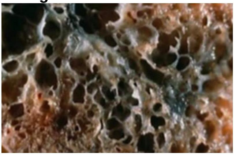
A 35-year-old woman with a long history of dyspnea, chronic cough, sputum production, and wheezing dies of respiratory failure following a bout of lobar pneumonia. She was not a smoker or an alcoholic. Which of the following underlying conditions is most likely associated with the pathologic changes shown in the lung autopsy?

Explanation: ***BAP1 loss*** - Biallelic **BAP1 inactivation**, detected as a loss of nuclear BAP1 immunoreactivity, is a highly specific marker for distinguishing **malignant mesothelioma** from benign reactive mesothelial proliferations. - While other markers confirm mesothelial lineage, only **BAP1 loss** directly points towards malignancy in this context. *Calretinin* - **Calretinin** is a sensitive marker for **mesothelial differentiation**, meaning it is expressed in both reactive and neoplastic mesothelial cells. - Therefore, it cannot differentiate between **benign reactive mesothelium** and **malignant mesothelioma**. *WT1* - **WT1 (Wilms Tumor 1)** is another valuable marker for **mesothelial lineage**, showing nuclear staining in both reactive and neoplastic mesothelial cells. - Like calretinin, its presence indicates mesothelial origin but does not distinguish between **benign and malignant processes**. *D2-40* - **D2-40 (podoplanin)** is a cell surface glycoprotein that is reliably expressed by normal and neoplastic mesothelial cells. - It is used to confirm the **mesothelial nature** of a proliferation but is not specific for malignancy.
Explanation: ***Streptococcus*** - After **E. coli**, **Streptococcus species** are the most common pathogens isolated in cases of **spontaneous bacterial peritonitis (SBP)**. - This includes various Streptococcal strains, which can translocate from the gut lumen into the ascetic fluid. *Bacteroids* - **Bacteroides fragilis** and other Bacteroids species are obligate anaerobes and are more commonly associated with **secondary peritonitis** due to bowel perforation, rather than SBP. - While they are abundant in the gut, their recovery in SBP is rare, suggesting a different pathogenic mechanism. *Enterococcus* - **Enterococci** are found in the gut flora and can cause SBP, but they are less common than Streptococcus species as the second leading cause. - Infections with Enterococcus are often seen in patients with **nosocomial infections** or those with prior antibiotic exposure. *Klebsiella* - **Klebsiella pneumoniae** is a common Gram-negative bacterium that can cause SBP, but it is typically the third most common Gram-negative cause after E. coli. - While significant, it does not surpass Streptococcus as the second most common overall cause of SBP following E. coli.
Explanation: ***Bronchopulmonary Aspergillosis*** - Allergic bronchopulmonary aspergillosis (ABPA) is a hypersensitivity reaction to *Aspergillus* species, primarily causing **bronchospasm**, **mucus plugging**, and **bronchiectasis**, rarely leading to pneumothorax [1]. - While it can cause significant lung damage, **pneumothorax** is not a characteristic or common complication, unlike the other conditions listed. *Marfan syndrome* - Patients with **Marfan syndrome** have connective tissue abnormalities, including those affecting the pleura and lung parenchyma. - This predisposition can lead to the formation of **apical blebs and bullae**, which are prone to rupture and cause **spontaneous pneumothorax** [2]. *Assisted ventilation* - **Positive pressure ventilation**, especially with high pressures or volumes, can cause barotrauma or volutrauma to the lungs [2]. - This can lead to alveolar rupture, resulting in **pneumothorax**, particularly in patients with pre-existing lung disease or those requiring high ventilatory support. *Eosinophilic granuloma* - Also known as **pulmonary Langerhans cell histiocytosis**, this condition involves the infiltration of the lungs by Langerhans cells. - It often leads to the formation of **cysts and nodules**, which can rupture and cause recurrent **spontaneous pneumothorax**.
Explanation: ***Pleural fluid ADA < 16*** - **Adenosine deaminase (ADA)** levels are used to diagnose **tuberculous pleural effusions**, with high levels (>40 U/L) suggesting exudate. [1] - A pleural fluid ADA of < 16 U/L is indicative of a **transudative effusion**, as it rules out tuberculosis. [1] *Pleural fluid LDH : serum LDH ratio > 0.6* - This criterion, where the ratio of **pleural fluid LDH** to **serum LDH** is greater than 0.6, is one of the classic **Light's criteria** for identifying an exudative effusion. [1] - An exudate typically has higher protein and enzyme content due to increased capillary permeability or local production. [1] *Pleural fluid protein : serum protein ratio > 0.5* - This indicates that the **protein concentration** in the pleural fluid is significantly higher than in the serum. [1] - This ratio is a key component of **Light's criteria** and suggests an inflammatory or exudative process. [1] *Pleural fluid LDH > two-thirds of the upper limit of serum LDH* - This is another major criterion in **Light's criteria** for defining an exudative pleural effusion. [1] - An elevated **pleural fluid LDH** suggests increased cellular activity or cell breakdown within the pleural space, characteristic of an exudate. [1]
Explanation: ***Parapneumonic effusion and Empyema*** - In **parapneumonic effusion** and **empyema**, bacteria and inflammatory cells consume glucose, leading to decreased pleural fluid glucose levels [1]. - A pleural fluid glucose level less than **60 mg/dL** or less than 50% of serum glucose is characteristic of these conditions. *Pulmonary embolism* - Pleural effusions associated with **pulmonary embolism** are typically transudates or exudates, but usually have normal glucose levels [1]. - The effusion results from increased **capillary permeability** or **venous hypertension**, not bacterial glucose consumption. *Pulmonary embolism and Empyema* - While **empyema** is correctly associated with low pleural glucose, **pulmonary embolism** is not. - Combining these conditions as a single answer is therefore inaccurate for the question asked. *Parapneumonic effusion* - This option is partially correct as **parapneumonic effusion** does cause decreased glucose [1]. - However, it is less comprehensive than the option that also includes **empyema**, which is a more severe form of parapneumonic effusion and also presents with low glucose [2].
Explanation: ***Malignancy*** - **Malignant pleural effusions** can be hemorrhagic due to tumor invasion of blood vessels and increased vascular fragility [1]. - Tumors frequently metastasize to the pleura, often leading to **hemothorax** due to friable microvasculature [1]. *Congestive heart failure* - Typically causes a **transudative pleural effusion** due to increased hydrostatic pressure, which is usually serous, not bloody [1]. - While fluid overload occurs, it does not directly lead to **blood accumulation** in the pleural space. *Myxoma* - A **benign cardiac tumor** that can embolize or cause constitutional symptoms, but it does not directly cause hemothorax. - Pleural effusions associated with myxomas are generally serous and related to **cardiac dysfunction**, not hemorrhage. *Rheumatoid arthritis* - Can cause **pleural effusions** (rheumatoid pleurisy), which are typically exudative and lymphocytosis-predominant [1]. - These effusions are rarely hemorrhagic and generally do not lead to **gross blood** in the pleural space.
Explanation: ***Asbestosis*** - Mesothelioma is a rare but aggressive cancer of the **pleura** or **peritoneum**, and its strongest known etiological link is with **asbestos exposure**. - Asbestos fibers can become lodged in the lungs and pleural lining, leading to chronic inflammation, DNA damage, and eventually oncogenic transformation. *Silicosis* - **Silicosis** is a lung disease caused by inhaling **crystalline silica dust**, primarily affecting miners, construction workers, and foundry workers. - While it can lead to pulmonary fibrosis and an increased risk of tuberculosis and lung cancer, it is not directly associated with mesothelioma. *Anthracosis* - **Anthracosis** is often seen in coal miners and urban dwellers due to the inhalation of **carbon dust**, leading to the accumulation of pigment in the lungs. - This condition is generally benign but can contribute to the development of **coal worker's pneumoconiosis**, which is distinct from mesothelioma. *Byssinosis* - **Byssinosis** is an occupational lung disease caused by the inhalation of **cotton dust** or other textile dusts, typically affecting textile workers. - Symptoms include chest tightness and shortness of breath, particularly after beginning work after a break, and it is unrelated to mesothelioma.
Explanation: ***Air in the pleural space*** - **Pneumothorax** is defined by the presence of **air in the pleural space**, which is the potential space between the parietal and visceral pleura [1]. - This accumulation of air causes a partial or complete **collapse of the lung** on the affected side due to the loss of negative intrapleural pressure [1]. *Cavity in the lung* - A **cavity in the lung** typically refers to a localized area of necrosis and excavation within the lung parenchyma, often seen in conditions like tuberculosis, fungal infections, or lung abscess. - While it can lead to pneumothorax if it ruptures into the pleural space, a lung cavity itself is not synonymous with pneumothorax. *Pus in the pleural space* - **Pus in the pleural space** is known as **empyema**, a type of pleural effusion characterized by purulent fluid accumulation due to infection [2]. - This is a distinct condition from pneumothorax, which involves air, not pus, in the pleural space. *Blood in the pleural space* - **Blood in the pleural space** is termed **hemothorax**, usually resulting from trauma, surgery, or underlying medical conditions that cause bleeding into the pleural cavity. - Hemothorax involves fluid (blood) accumulation, whereas pneumothorax specifically involves air.
Explanation: ***Asbestosis*** - Ferruginous bodies are specifically associated with **exposure to asbestos**, which leads to asbestosis [1]. - These bodies are seen as **siderophilic structures** resembling a "dumbbell" shape under the microscope, which are indicative of this condition [1]. *Bagassosis* - Caused by exposure to **bagasse dust**, primarily from sugarcane, leading to allergic alveolitis rather than ferruginous bodies [1]. - Histopathology typically shows **lymphocytic infiltration** and non-caseating granulomas, not ferruginous bodies. *Byssinosis* - This is associated with inhalation of **cotton dust** and primarily results in **bronchoconstriction** and respiratory symptoms rather than ferruginous bodies. - Characterized by a **respiratory illness** that worsens at the beginning of the work week, missing the key features of asbestosis. *Silicosis* - Resulting from exposure to **silica dust**, this condition leads to macules and nodules in the lungs instead of ferruginous bodies [1]. - The hallmark findings are **hyaline nodules** on imaging and not the abnormal iron-containing structures seen in asbestosis. **References:** [1] Kumar V, Abbas AK, et al.. Robbins and Cotran Pathologic Basis of Disease. 9th ed. The Lung, pp. 695, 698-699.
Explanation: ***Alpha 1 antitrypsin deficiency*** - This condition leads to **accumulation of abnormal protein** in the liver and lungs, resulting in emphysema, which is consistent with chronic cough and dyspnea [1]. - Patients often develop **lung pathology** similar to what is seen in smokers, making it plausible given the patient's background [1]. *Mutation in dynein arms* - This is associated with **primary ciliary dyskinesia**, which presents with recurrent respiratory infections but is not typical in non-smokers or in the context of **dyspnea with chronic cough**. - Usually linked to **situs inversus** and **recurrent infections**, neither of which is highlighted here. *Antibodies against type 4 collagen* - This condition is related to **Goodpasture syndrome**, which typically results in **hemoptysis** and **renal failure**, rather than chronic cough and sputum production. - The predominant involvement in this syndrome does not align with the clinical presentation of **chronic lung disease** noted in this patient. *Cystic fibrosis* - While it causes **chronic respiratory symptoms**, it is usually seen in younger patients and is associated with **pancreatic insufficiency** and **salty sweat**. - The age of the patient and symptom progression does not fit well with a diagnosis of cystic fibrosis. **References:** [1] Kumar V, Abbas AK, et al.. Robbins and Cotran Pathologic Basis of Disease. 9th ed. The Lung, pp. 683-684.
Get full access to all questions, explanations, and performance tracking.
Start For Free