
Respiratory Pathology — MCQs

On this page
A lung section from an autopsy of an AIDS patient with a CD4 count less than 100/mm³ shows desquamation of type 1 pneumocytes with prominent intranuclear basophilic inclusion bodies surrounded by a clear halo. What is the most likely diagnosis causing these features?
All of the following statements are true regarding the histological features of emphysema, except?
Asbestosis of the lung is associated with all of the following except?
Which of the following is the most characteristic feature of Adult Respiratory Distress Syndrome (ARDS)?
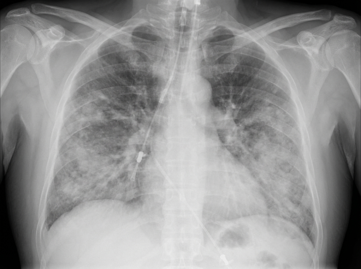
What is the pathophysiological cause of the disease shown in the X-ray and gross specimen from an ICU patient?
The term "brown induration" is used in relation to which of the following organs?
In the course of a laryngoscopic examination for hoarseness, a small lesion is found on the true vocal cord of a 57-year-old male smoker. On biopsy, severe squamous dysplasia is noted. If untreated, this lesion may progress to which of the following?
A patient presents with a stony hard, painless lymph node in the left supraclavicular fossa. A biopsy report states squamous cell carcinoma. What is the most likely primary diagnosis?
Rasmussen's aneurysm is seen in which of the following arteries?
BAL of a patient shows foamy macrophages with decreased CD4:CD8 ratio. What is the diagnosis?
Practice by Chapter
Congenital Anomalies
Practice Questions
Atelectasis and Acute Lung Injury
Practice Questions
Obstructive Pulmonary Diseases
Practice Questions
Restrictive Pulmonary Diseases
Practice Questions
Lung Infections
Practice Questions
Pulmonary Vascular Diseases
Practice Questions
Lung Tumors
Practice Questions
Pleural Diseases
Practice Questions
Interstitial Lung Diseases
Practice Questions
Occupational Lung Diseases
Practice Questions
Want unlimited practice?
Get full access to all questions, explanations, and performance tracking.
Scan to download app