A 60-year-old man presents with chronic obstructive pulmonary disease (COPD). Which type of emphysema is most commonly associated with smoking?
A 55-year-old male with a history of chronic bronchitis is being evaluated for respiratory distress. Which pathophysiological process is primarily involved in chronic bronchitis?
A 65-year-old woman presents with progressive dyspnea and a productive cough. A CT scan reveals a lung mass and pleural effusion. A biopsy shows adenocarcinoma. Which of the following is a common histologic feature?
In RDS in a child, which cells are found defective?
Anthracosis is due to inhalation of?
Which of the following is a characteristic feature of chronic bronchitis?
Which of the following statements about silicosis is true?
Which type of pneumoconiosis is most commonly associated with tuberculosis (TB)?
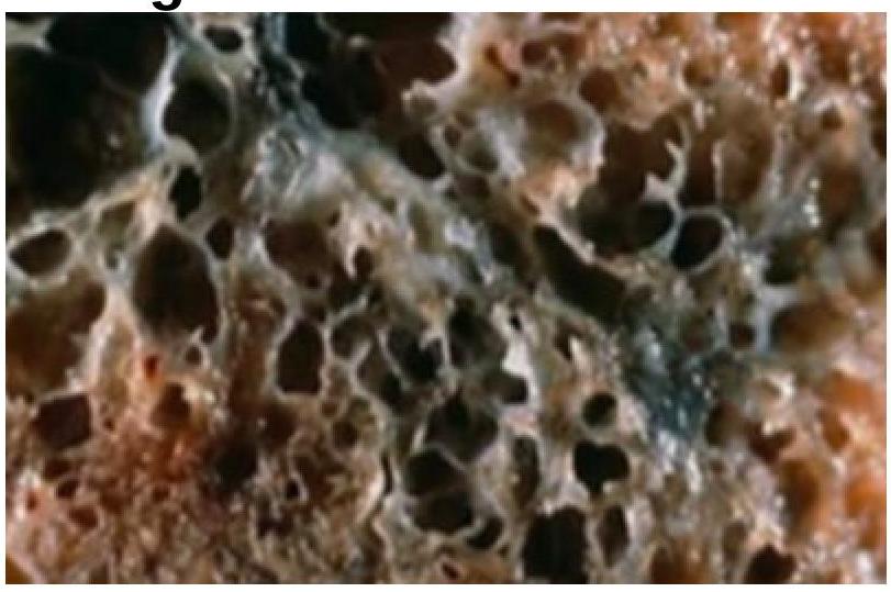
A 35-year-old woman with a long history of dyspnea, chronic cough, sputum production, and wheezing dies of respiratory failure following a bout of lobar pneumonia. She was not a smoker or an alcoholic. Which of the following underlying conditions is most likely associated with the pathologic changes shown in the lung autopsy?

Most common cause of idiopathic interstitial pneumonia is
Explanation: ***Centriacinar emphysema*** - It is the **most common type associated with smoking** [1], particularly in the upper lobes of the lungs. - Characterized by **destruction of the respiratory bronchioles**, leading to airflow obstruction in COPD patients [2]. *Irregular emphysema* - Often seen in older patients and is usually **associated with scarring** from various lung diseases. - It does not have a clear link to **smoking** like centriacinar emphysema does. *Paraseptal emphysema* - Primarily affects the **distal alveoli** and is often associated with **spontaneous pneumothorax**, not smoking. - Less common in COPD patients and more related to **lung apices** than to smoking. *Panacinar emphysema* - Typically linked to **Alpha-1 Antitrypsin deficiency** [1], not primarily to smoking. - It causes uniform enlargement of the acini, predominantly affecting the **lower lobes**. **References:** [1] Kumar V, Abbas AK, et al.. Robbins and Cotran Pathologic Basis of Disease. 9th ed. The Lung, pp. 684-685. [2] Cross SS. Underwood's Pathology: A Clinical Approach. 6th ed. Common Clinical Problems From Respiratory Tract Disease, pp. 326-327.
Explanation: ***Airway inflammation and obstruction*** - Chronic bronchitis is characterized by **persistent inflammation** of the airways, leading to **mucous hypersecretion** and obstruction [1]. - This results in **increased airway resistance** and respiratory distress, essential in understanding the pathophysiology [1]. *Alveolar collapse* - Alveolar collapse occurs primarily in conditions like **atelectasis** and is not a primary feature of chronic bronchitis. - Chronic bronchitis mainly affects the **bronchi and bronchioles**, not directly causing alveolar issues [1]. *Increased pulmonary surfactant production* - Surfactant is primarily involved in **alveolar stability**; its production does not increase in chronic bronchitis. - In fact, surfactant levels may be normal or decreased, as the disease focuses on airway obstruction rather than alveolar dynamics. *Pulmonary fibrosis* - Pulmonary fibrosis involves **scarring of lung tissue**, primarily affecting the interstitium, not the airway-centric pathology seen in chronic bronchitis. - While chronic bronchitis may arise from long-standing lung irritation, it typically does not lead to **fibrosing patterns** as seen in interstitial lung diseases. **References:** [1] Kumar V, Abbas AK, et al.. Robbins and Cotran Pathologic Basis of Disease. 9th ed. The Lung, pp. 685-686.
Explanation: ***Glandular formation with mucin production*** - Adenocarcinoma of the lung frequently exhibits **glandular structures** that secrete mucin, a hallmark of this carcinoma subtype. - This feature aids in distinguishing it from other lung cancer types, emphasizing its **secretory nature**. *Squamous differentiation* - This feature is typical of **squamous cell carcinoma**, not adenocarcinoma, which arises from **glandular epithelium**. - Presence of keratinization and **intercellular bridges** are more characteristic of squamous differentiation. *Neuroendocrine features* - Neuroendocrine features are more associated with **small cell lung cancer** and other neuroendocrine tumors, not with adenocarcinoma [1]. - Adenocarcinoma typically lacks **neuroendocrine differentiation**, focusing instead on glandular characteristics. *Keratin pearls* - Keratin pearls are indicative of **squamous cell carcinoma**, which involves keratinization and does not occur in adenocarcinoma. - The formation of these pearls is a sign of **differentiation towards squamous features**, absent in glandular tissue. **References:** [1] Cross SS. Underwood's Pathology: A Clinical Approach. 6th ed. Common Clinical Problems From Respiratory Tract Disease, pp. 337-338.
Explanation: ***Type 2 pneumocytes*** - **Type 2 pneumocytes** are responsible for producing **surfactant**, which reduces surface tension in the alveoli and prevents their collapse [2], [3]. - In **Respiratory Distress Syndrome (RDS)**, especially in premature infants, these cells are immature or insufficient, leading to **surfactant deficiency** [1]. - This is the **primary cellular defect** in RDS, making it the correct answer [2]. *Bronchial epithelium* - The **bronchial epithelium** primarily lines the conducting airways and is involved in mucociliary clearance, not surfactant production. - Defects in this epithelium are associated with conditions like **bronchitis** or **asthma**, not the primary pathogenesis of RDS. *Type 1 pneumocytes* - **Type 1 pneumocytes** are thin, flat cells that form the majority of the alveolar surface (95%) and are crucial for gas exchange. - While they are affected by the damage in RDS, their primary defect is not the cause of the disease; the underlying problem is **surfactant deficiency** from Type 2 pneumocytes [2]. *Clara cells* - **Clara cells** (now called club cells) are non-ciliated secretory cells found in the bronchioles that produce Clara cell secretory protein (CCSP). - They play a role in detoxification and immune modulation but are **not responsible for surfactant production** or the pathogenesis of RDS. **References:** [1] Kumar V, Abbas AK, et al.. Robbins and Cotran Pathologic Basis of Disease. 9th ed. Diseases of Infancy and Childhood, pp. 465-466. [2] Cross SS. Underwood's Pathology: A Clinical Approach. 6th ed. Common Clinical Problems From Respiratory Tract Disease, pp. 313-314. [3] Kumar V, Abbas AK, et al.. Robbins and Cotran Pathologic Basis of Disease. 9th ed. Diseases of Infancy and Childhood, p. 466.
Explanation: ***Coal dust*** - Anthracosis is primarily caused by the inhalation of **coal dust**, leading to carbon accumulation in the lungs [1]. - This condition is commonly associated with **coal miners** and is a type of **pneumoconiosis** [1]. *Asbestos* - Inhalation of **asbestos fibers** leads to conditions like **asbestosis** and **mesothelioma**, not anthracosis [1]. - Symptoms include **lung fibrosis** and are characterized by a different pathophysiological pattern. *Beryllium dust* - Exposure to **beryllium dust** can cause **berylliosis**, which manifests as a chronic lung disease involving granuloma formation [1]. - It is a distinct condition and does not relate to the carbon deposits seen in anthracosis. *Silica dust* - Inhalation of **silica dust** causes **silicosis**, characterized by nodular lung disease and is distinct from anthracosis [1]. - Silicosis is associated with **mining** and **stone cutting**, not coal dust inhalation. **References:** [1] Kumar V, Abbas AK, et al.. Robbins and Cotran Pathologic Basis of Disease. 9th ed. The Lung, p. 695.
Explanation: ***Bronchiolitis obliterans*** - Chronic bronchitis is often associated with ends of the bronchi, leading to **peripheral airway obstruction**, which may evolve into bronchiolitis obliterans [1]. - It manifests as progressive **dyspnea** and chronic cough due to significant **airway inflammation** and damage [1]. *Decrease in Reid index* - The Reid index (ratio of gland size to airway size) typically **increases** in chronic bronchitis due to mucous gland hyperplasia. - A decrease would suggest a **shift towards airway obstruction** rather than gland enlargement. *Increase in number of glands without any change in size* - Chronic bronchitis involves an **increase in both number and size** of mucous glands, as seen in the Reid index. - This oes not accurately reflect the **pathological changes** that occur in this condition. *Only large airways are involved* - Chronic bronchitis affects both **large and small airways**, including the terminal bronchioles [1]. - This misrepresents the widespread impact of the disease on the **entire bronchial tree**, not limited to larger airways. **References:** [1] Kumar V, Abbas AK, et al.. Robbins and Cotran Pathologic Basis of Disease. 9th ed. The Lung, pp. 685-686.
Explanation: ***Associated with tuberculosis*** - Silicosis significantly increases the risk of developing **tuberculosis** due to silicate dust impairing immune function in the lungs [1]. - There is a clear **epidemiological link** between silicosis and the incidence of pulmonary tuberculosis [1]. *Produces pleural plaque* - Pleural plaques are more characteristic of **asbestosis**, not silicosis [3]; silicosis typically involves **nodular interstitial fibrosis** [2]. - Silicosis primarily leads to **nodules** in the upper lung zones rather than pleural changes like plaques [2]. *Lower lobe infiltration* - Silicosis typically shows **upper lobe** involvement with nodular opacities rather than lower lobe infiltrations seen in other diseases like asbestosis or pulmonary edema [2]. - The lung radiology in silicosis is distinct, characterized by **upper lobe fibrosis** rather than localized lower lobe changes. *All* - Since only one option regarding silicosis is true, this blanket statement lacks accuracy; not all statements listed are correct. - Each detail must be evaluated individually, and not all represent true aspects of silicosis, making "all" an incorrect conclusion. **References:** [1] Kumar V, Abbas AK, et al.. Robbins and Cotran Pathologic Basis of Disease. 9th ed. The Lung, pp. 697-698. [2] Kumar V, Abbas AK, et al.. Robbins and Cotran Pathologic Basis of Disease. 9th ed. The Lung, p. 697. [3] Kumar V, Abbas AK, et al.. Robbins and Cotran Pathologic Basis of Disease. 9th ed. The Lung, p. 695.
Explanation: ***Silicosis*** - The most common type of pneumoconiosis, **silicosis**, is specifically associated with an increased risk of developing **tuberculosis (TB)** due to the immune suppression caused by silica dust exposure [1]. - Characterized by **reticular opacities** and **nodules** on chest imaging, it leads to progressive lung impairment and predisposes patients to TB reactivation. *Bysinosis* - Caused by exposure to **cotton dust**, leading to respiratory symptoms, it is not closely linked with TB. - Primarily affects **textile workers** and presents with symptoms like shortness of breath but lacks the association with **lung fibrosis** or TB risks seen in silicosis. *Asbestosis* - Resulting from asbestos exposure, it leads to **interstitial fibrosis** and is primarily associated with lung cancer and mesothelioma, rather than TB. - While it causes significant respiratory issues, it does not have the same predisposition to TB as seen with silicosis. *Baggassosis* - A less common type of pneumoconiosis caused by inhalation of **sugarcane dust**, leading to respiratory problems but is not associated with TB. - It primarily affects **agricultural workers** and involves respiratory symptoms but lacks the defining features relevant to TB risk. **References:** [1] Kumar V, Abbas AK, et al.. Robbins and Cotran Pathologic Basis of Disease. 9th ed. The Lung, pp. 697-698.
Explanation: ***Alpha 1 antitrypsin deficiency*** - This condition leads to **accumulation of abnormal protein** in the liver and lungs, resulting in emphysema, which is consistent with chronic cough and dyspnea [1]. - Patients often develop **lung pathology** similar to what is seen in smokers, making it plausible given the patient's background [1]. *Mutation in dynein arms* - This is associated with **primary ciliary dyskinesia**, which presents with recurrent respiratory infections but is not typical in non-smokers or in the context of **dyspnea with chronic cough**. - Usually linked to **situs inversus** and **recurrent infections**, neither of which is highlighted here. *Antibodies against type 4 collagen* - This condition is related to **Goodpasture syndrome**, which typically results in **hemoptysis** and **renal failure**, rather than chronic cough and sputum production. - The predominant involvement in this syndrome does not align with the clinical presentation of **chronic lung disease** noted in this patient. *Cystic fibrosis* - While it causes **chronic respiratory symptoms**, it is usually seen in younger patients and is associated with **pancreatic insufficiency** and **salty sweat**. - The age of the patient and symptom progression does not fit well with a diagnosis of cystic fibrosis. **References:** [1] Kumar V, Abbas AK, et al.. Robbins and Cotran Pathologic Basis of Disease. 9th ed. The Lung, pp. 683-684.
Explanation: ***Idiopathic pulmonary fibrosis (IPF)*** - This is the **most common** form of idiopathic interstitial pneumonia, accounting for approximately **50-60% of all IIP cases** - Represents the **most severe** IIP subtype with poor prognosis - Characterized by progressive **usual interstitial pneumonia (UIP) pattern** with fibroblastic foci and honeycombing - Presents with progressive dyspnea, dry cough, and restrictive lung disease *Organizing pneumonia* - While **Cryptogenic Organizing Pneumonia (COP)** is a form of idiopathic interstitial pneumonia, it is **much less common than IPF** [1] - Characterized by **intra-alveolar granulation tissue (Masson bodies)** [1] - Better prognosis and steroid-responsive compared to IPF [1] *Sarcoidosis* - This is **NOT classified as an idiopathic interstitial pneumonia** - It is a separate **multisystem granulomatous disease** with **non-caseating granulomas** - Has a distinct etiology related to altered immune response - Does not belong to the IIP classification system *Lipoid pneumonia* - This is **NOT an idiopathic interstitial pneumonia** - Results from **aspiration of lipid substances** causing exogenous lipoid pneumonia - Has a **known extrinsic cause**, therefore not "idiopathic" - Not part of the IIP classification **References:** [1] Cross SS. Underwood's Pathology: A Clinical Approach. 6th ed. Common Clinical Problems From Respiratory Tract Disease, pp. 330-331.
Congenital Anomalies
Practice Questions
Atelectasis and Acute Lung Injury
Practice Questions
Obstructive Pulmonary Diseases
Practice Questions
Restrictive Pulmonary Diseases
Practice Questions
Lung Infections
Practice Questions
Pulmonary Vascular Diseases
Practice Questions
Lung Tumors
Practice Questions
Pleural Diseases
Practice Questions
Interstitial Lung Diseases
Practice Questions
Occupational Lung Diseases
Practice Questions
Get full access to all questions, explanations, and performance tracking.
Start For Free