
Respiratory Pathology — MCQs

On this page
In RDS in a child, which cells are found defective?
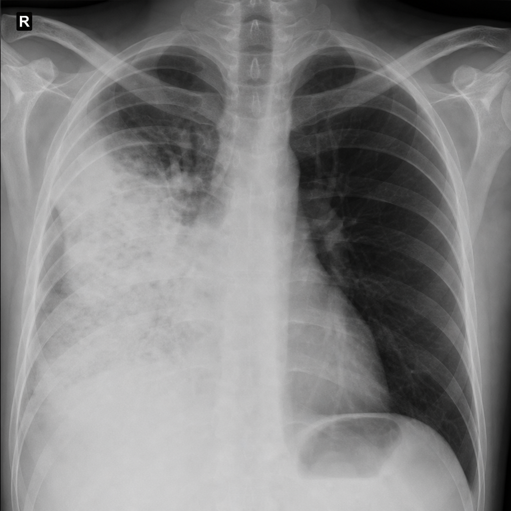
Identify the condition shown in the image of the lung.
Granulomatous lung disease is caused by?
Which of the following statements about silicosis is true?
Which of the following is a characteristic feature of chronic bronchitis?
Anthracosis is due to inhalation of?
Which type of pneumoconiosis is most commonly associated with tuberculosis (TB)?
Metastasis to lungs comes most commonly from
In which type of lung carcinoma is the p53 mutation most commonly observed?
In which condition is Calretinin primarily used as a diagnostic marker?
Practice by Chapter
Congenital Anomalies
Practice Questions
Atelectasis and Acute Lung Injury
Practice Questions
Obstructive Pulmonary Diseases
Practice Questions
Restrictive Pulmonary Diseases
Practice Questions
Lung Infections
Practice Questions
Pulmonary Vascular Diseases
Practice Questions
Lung Tumors
Practice Questions
Pleural Diseases
Practice Questions
Interstitial Lung Diseases
Practice Questions
Occupational Lung Diseases
Practice Questions
Want unlimited practice?
Get full access to all questions, explanations, and performance tracking.
Scan to download app