
Respiratory Pathology — MCQs

On this page
A 70-year-old smoker presents with chronic cough and hypoxia. What is the primary pathophysiological change in the alveoli contributing to hypoxia in emphysema?
A 52-year-old male with a history of smoking presents with a persistent cough and hemoptysis. A CT scan reveals a central lung mass, and a biopsy shows small, round, blue cells with neuroendocrine differentiation. What is the most likely diagnosis?
A 60-year-old man presents with chronic obstructive pulmonary disease (COPD). Which type of emphysema is most commonly associated with smoking?
A 55-year-old male with a history of chronic bronchitis is being evaluated for respiratory distress. Which pathophysiological process is primarily involved in chronic bronchitis?
A 28-year-old woman presents with shortness of breath and cough. A chest X-ray reveals multiple bilateral lung nodules. A biopsy shows multinucleated giant cells and asteroid bodies. What is the most likely diagnosis?
In a 60-year-old female with a smoking history presenting with chronic cough, hemoptysis, and weight loss, how can immunohistochemical markers be used to differentiate between squamous cell carcinoma and small cell lung cancer?
Which neoplasm is most strongly associated with asbestos exposure?
A 65-year-old woman presents with progressive dyspnea and a productive cough. A CT scan reveals a lung mass and pleural effusion. A biopsy shows adenocarcinoma. Which of the following is a common histologic feature?
A 60-year-old male presents with chronic cough and hemoptysis. Bronchoscopy reveals a mass obstructing the left main bronchus. Which lymph nodes are most likely to show metastasis first?
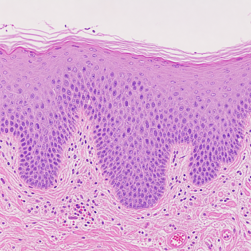
A 45-year-old man who is a chronic smoker came to the clinic with a complaint of cough. The physician examines the patient and takes a biopsy. The biopsy image is provided below. Which of the following cellular changes has happened to this patient?
Practice by Chapter
Congenital Anomalies
Practice Questions
Atelectasis and Acute Lung Injury
Practice Questions
Obstructive Pulmonary Diseases
Practice Questions
Restrictive Pulmonary Diseases
Practice Questions
Lung Infections
Practice Questions
Pulmonary Vascular Diseases
Practice Questions
Lung Tumors
Practice Questions
Pleural Diseases
Practice Questions
Interstitial Lung Diseases
Practice Questions
Occupational Lung Diseases
Practice Questions
Want unlimited practice?
Get full access to all questions, explanations, and performance tracking.
Scan to download app