
Respiratory Pathology — MCQs

On this page
Carcinoid tumors of the lung arise from which of the following cells?
Which of the following statements regarding oat cell carcinoma of the lung is FALSE?
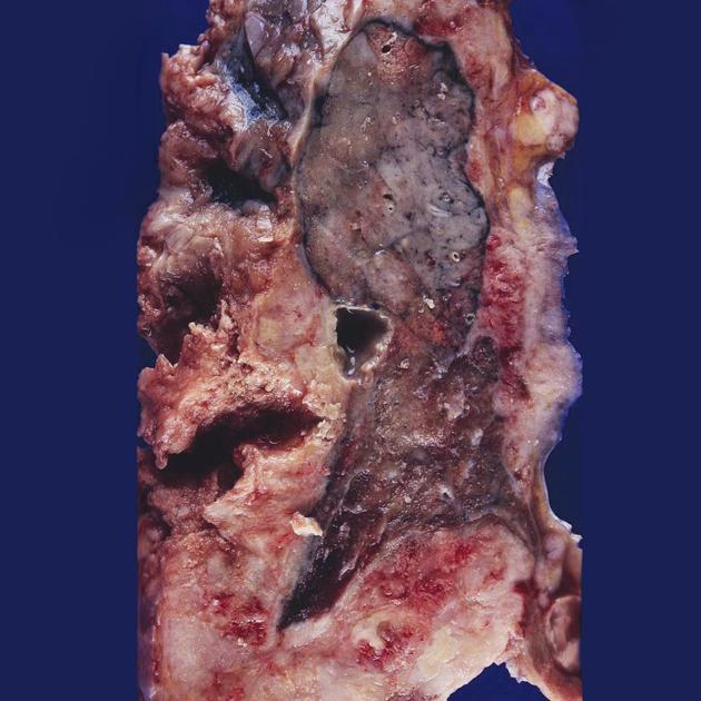
A 40-year-old female presented with a 3-month history of fever, loss of appetite, weight loss, cough, and weakness. A chest X-ray revealed consolidation in the upper lung. She died a few days after admission. Autopsy revealed gross lung findings and biopsy findings. What is your diagnosis?
Caplan syndrome is seen in which of the following conditions?
Which type of lung carcinoma is most commonly associated with causing Superior Vena Cava (SVC) syndrome?
Ghons focus is seen in?
Which of the following is NOT a typical histological feature of Hypersensitivity Pneumonitis?
Which of the following statements about lung carcinoma is true?
Which of the following is true about bronchial carcinoids?
What condition is indicated by the presence of Creola bodies in sputum?
Practice by Chapter
Congenital Anomalies
Practice Questions
Atelectasis and Acute Lung Injury
Practice Questions
Obstructive Pulmonary Diseases
Practice Questions
Restrictive Pulmonary Diseases
Practice Questions
Lung Infections
Practice Questions
Pulmonary Vascular Diseases
Practice Questions
Lung Tumors
Practice Questions
Pleural Diseases
Practice Questions
Interstitial Lung Diseases
Practice Questions
Occupational Lung Diseases
Practice Questions
Want unlimited practice?
Get full access to all questions, explanations, and performance tracking.
Scan to download app