
Respiratory Pathology — MCQs

On this page
A 67-year-old male with a history of chronic smoking presents with hemoptysis and cough. Bronchoscopic biopsy from a centrally located mass shows an undifferentiated tumor histopathologically. Which immunohistochemical marker would be most useful to make a proper diagnosis?
A 45-year-old man incurs blunt chest trauma. On examination, he has marked right chest wall pain. A chest radiograph shows a fractured 7th rib on the right side. Over the next 2 days, he has subcutaneous soft tissue swelling with nonpainful crepitus on palpation of the right chest wall. Leakage of which of the following is most likely producing this swelling?
A patient presents with a severe form of atopic asthma. Which of the following changes would most likely be found in this patient's blood?
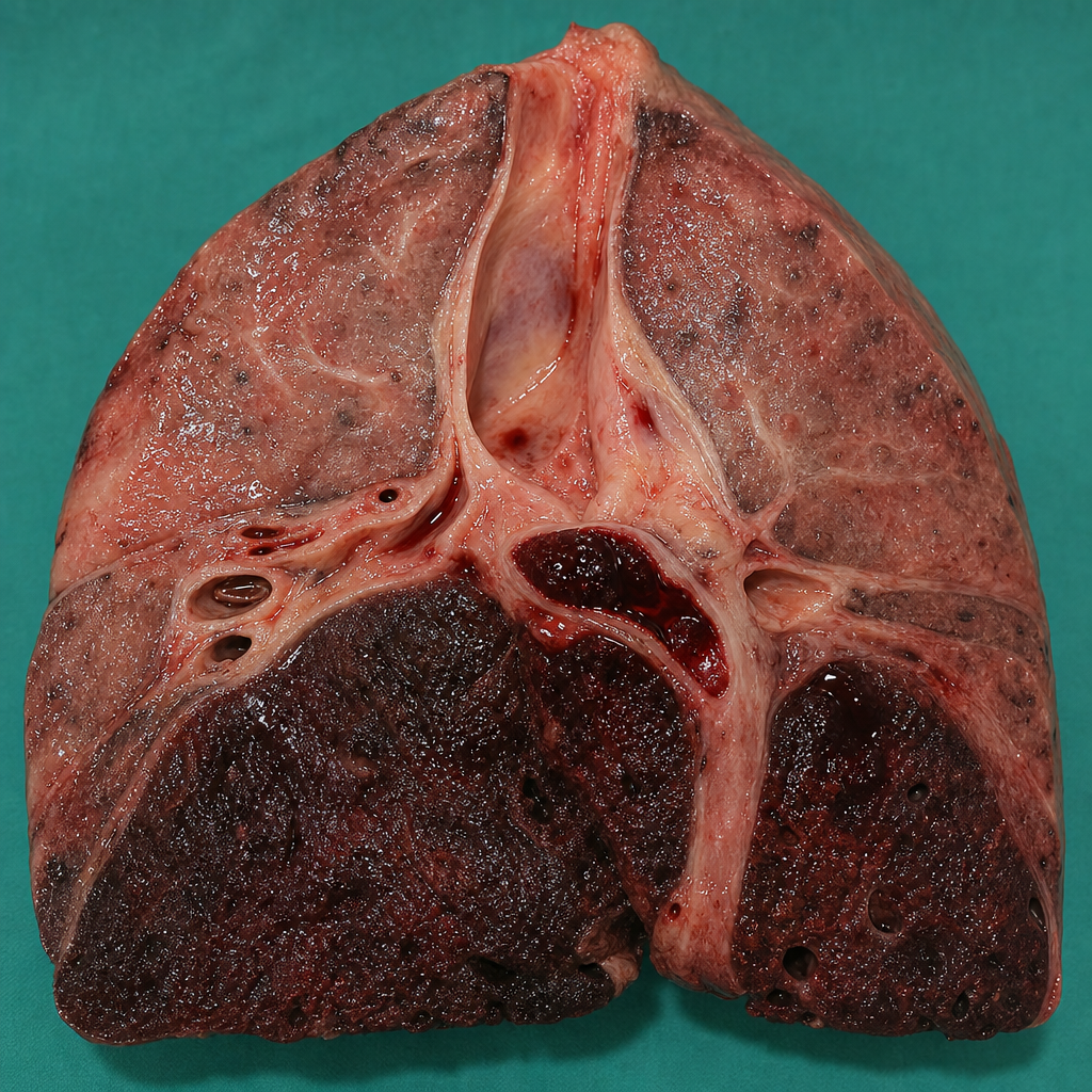
A 22-year-old biker sustained severe injuries in a road traffic accident. After admission to the hospital, his condition suddenly worsened, leading to death. A pathological specimen is provided. What is the likely cause of death?
What is the characteristic pathological feature of pneumococcal pneumonia?
Which type of lung tumour responds best to radiotherapy?
A 50-year-old man with a neurodegenerative disease presents with fever and cough productive of yellow sputum for 3 days. Physical examination reveals dullness to percussion at the left lung base. A chest radiograph shows consolidation in the left lower lobe. The patient develops an abscess despite antibiotic therapy and subsequently dies. Autopsy reveals a bronchopleural fistula surrounded by a pronounced fibroblastic reaction. Gross examination of the abscess shows small, yellow, 1-cm to 2-mm "sulfur granules." Which of the following organisms is most likely to produce these findings?
What is true regarding adenocarcinoma of the lung?
What is the most common source of pulmonary embolism?
Giant cell (Hecht's) pneumonia is typically associated with which of the following viruses?
Practice by Chapter
Congenital Anomalies
Practice Questions
Atelectasis and Acute Lung Injury
Practice Questions
Obstructive Pulmonary Diseases
Practice Questions
Restrictive Pulmonary Diseases
Practice Questions
Lung Infections
Practice Questions
Pulmonary Vascular Diseases
Practice Questions
Lung Tumors
Practice Questions
Pleural Diseases
Practice Questions
Interstitial Lung Diseases
Practice Questions
Occupational Lung Diseases
Practice Questions
Want unlimited practice?
Get full access to all questions, explanations, and performance tracking.
Scan to download app